 Movie
Movie Controller

[English] 日本語
 Yorodumi
- PDB-4wo8: The substrate-free duplicated taurocyamine kinase from Schistosom... -
+ Open data

- Basic information
| Entry | Database: PDB / ID: 4wo8 | ||||||
|---|---|---|---|---|---|---|---|
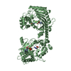
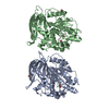
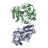
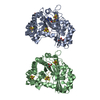
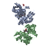
| Title | The substrate-free duplicated taurocyamine kinase from Schistosoma mansoni | ||||||
 Components Components | Taurocyamine kinase | ||||||
 Keywords Keywords | TRANSFERASE / duplicated / substrate specificity / transition state | ||||||
| Function / homology |  Function and homology information Function and homology informationtaurocyamine kinase / taurocyamine kinase activity / arginine kinase activity / phosphocreatine biosynthetic process / creatine kinase activity / oxidoreductase activity / : / ATP binding / metal ion binding Similarity search - Function | ||||||
| Biological species |  | ||||||
| Method |  X-RAY DIFFRACTION / SYNCHROTRON / MOLECULAR REPLACEMENT / Resolution: 2.2 Å X-RAY DIFFRACTION / SYNCHROTRON / MOLECULAR REPLACEMENT / Resolution: 2.2 Å | ||||||
 Authors Authors | Merceron, R. / Awama, A. / Montserret, R. / Marcillat, O. / Gouet, P. | ||||||
 Citation Citation | Journal: J.Biol.Chem. / Year: 2015 Title: The Substrate-free and -bound Crystal Structures of the Duplicated Taurocyamine Kinase from the Human Parasite Schistosoma mansoni. Authors: Merceron, R. / Awama, A.M. / Montserret, R. / Marcillat, O. / Gouet, P. | ||||||
| History |
|
- Structure visualization
| Structure viewer | Molecule: MolmilJmol/JSmol |
|---|
- Downloads & links
-Download
| PDBx/mmCIF format | 4wo8.cif.gz | 157.6 KB | Display | PDBx/mmCIF format |
|---|---|---|---|---|
| PDB format | pdb4wo8.ent.gz | 122.1 KB | Display | PDB format |
| PDBx/mmJSON format | 4wo8.json.gz | Tree view | PDBx/mmJSON format | |
| Others |  Other downloads Other downloads |
-Validation report
| Arichive directory | https://data.pdbj.org/pub/pdb/validation_reports/wo/4wo8ftp://data.pdbj.org/pub/pdb/validation_reports/wo/4wo8 | HTTPS FTP |
|---|
-Related structure data
| Related structure data | 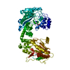 4wodC  4woeC 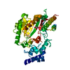 2j1qS S: Starting model for refinement C: citing same article ( |
|---|---|
| Similar structure data |


-Links
 PDBj
PDBj
- Assembly
| Deposited unit | 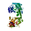
| ||||||||
|---|---|---|---|---|---|---|---|---|---|
| 1 |
| ||||||||
| Unit cell |
|
-Components
| #1: Protein | Mass: 80422.703 Da / Num. of mol.: 1 / Fragment: UNP residues 31-746 / Mutation: A11V D235G I237T D493G Source method: isolated from a genetically manipulated source Source: (gene. exp.)  |
|---|---|
| #2: Water | ChemComp-HOH /  Mass: 18.015 Da / Num. of mol.: 362 / Source method: isolated from a natural source / Formula: H2O Mass: 18.015 Da / Num. of mol.: 362 / Source method: isolated from a natural source / Formula: H2O |
-Experimental details
-Experiment
| Experiment | Method: X-RAY DIFFRACTION |
|---|
- Sample preparation
| Crystal | Density Matthews: 2.4 Å3/Da / Density % sol: 48.73 % |
|---|---|
| Crystal grow | Temperature: 293 K / Method: vapor diffusion, hanging drop / pH: 5.4 Details: 200 mM ammonium tartrate dibasic pH 5.4, 20% (w/v) polyethylene glycol 3350, 20% (v/v) ethylene glycol PH range: 5.4 - 5.6 |
-Data collection
| Diffraction | Mean temperature: 100 K |
|---|---|
| Diffraction source | Source: SYNCHROTRON / Site: SLS  / Beamline: X06DA / Wavelength: 1 Å / Beamline: X06DA / Wavelength: 1 Å |
| Detector | Type: DECTRIS PILATUS 2M / Detector: PIXEL / Date: Oct 13, 2013 |
| Radiation | Protocol: SINGLE WAVELENGTH / Monochromatic (M) / Laue (L): M / Scattering type: x-ray |
| Radiation wavelength | Wavelength: 1 Å / Relative weight: 1 |
| Reflection | Resolution: 2.2→50 Å / Num. all: 38515 / Num. obs: 38030 / % possible obs: 98.7 % / Redundancy: 3.1 % / Net I/σ(I): 11.71 |
| Reflection shell | Resolution: 2.2→2.26 Å / Redundancy: 2.2 % / Mean I/σ(I) obs: 2.11 / % possible all: 97 |
- Processing
| Software |
| |||||||||||||||||||||||||||||||||||||||||||||||||||||||||||||||||||||||||||||||||||||||||||||||||||||||||
|---|---|---|---|---|---|---|---|---|---|---|---|---|---|---|---|---|---|---|---|---|---|---|---|---|---|---|---|---|---|---|---|---|---|---|---|---|---|---|---|---|---|---|---|---|---|---|---|---|---|---|---|---|---|---|---|---|---|---|---|---|---|---|---|---|---|---|---|---|---|---|---|---|---|---|---|---|---|---|---|---|---|---|---|---|---|---|---|---|---|---|---|---|---|---|---|---|---|---|---|---|---|---|---|---|---|---|
| Refinement | Method to determine structure: MOLECULAR REPLACEMENT Starting model: 2j1q Resolution: 2.2→46.182 Å / SU ML: 0.23 / Cross valid method: FREE R-VALUE / σ(F): 1.36 / Phase error: 23.18 / Stereochemistry target values: ML
| |||||||||||||||||||||||||||||||||||||||||||||||||||||||||||||||||||||||||||||||||||||||||||||||||||||||||
| Solvent computation | Shrinkage radii: 0.9 Å / VDW probe radii: 1.11 Å / Solvent model: FLAT BULK SOLVENT MODEL | |||||||||||||||||||||||||||||||||||||||||||||||||||||||||||||||||||||||||||||||||||||||||||||||||||||||||
| Refinement step | Cycle: LAST / Resolution: 2.2→46.182 Å
| |||||||||||||||||||||||||||||||||||||||||||||||||||||||||||||||||||||||||||||||||||||||||||||||||||||||||
| Refine LS restraints |
| |||||||||||||||||||||||||||||||||||||||||||||||||||||||||||||||||||||||||||||||||||||||||||||||||||||||||
| LS refinement shell |
|
