Mass: 18.015 Da / Num. of mol.: 166 / Source method: isolated from a natural source / Formula: H2O
Sequence details
TWO RESIDUES HAVE BEEN MODIFIED BASED ON THE ELECTRON DENSITY MAP, LEU82 AND SER104. IN THE GENBANK ...TWO RESIDUES HAVE BEEN MODIFIED BASED ON THE ELECTRON DENSITY MAP, LEU82 AND SER104. IN THE GENBANK SEQUENCE, BOTH POSITIONS APPEAR AS PHE.
-
Experimental details
-
Experiment
Experiment
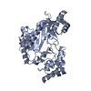
Method: X-RAY DIFFRACTION / Number of used crystals: 1
-
Sample preparation
Crystal
Density Matthews: 3.7 Å3/Da / Density % sol: 67 %
Crystal grow
pH: 7.5 / Details: 2.5 M AMMONIUM SULFATE, 0.1 M TRIS HCL, PH 7.5
Resolution: 1.9→75.16 Å / Cor.coef. Fo:Fc: 0.954 / Cor.coef. Fo:Fc free: 0.943 / SU B: 5.471 / SU ML: 0.084 / TLS residual ADP flag: LIKELY RESIDUAL / Cross valid method: THROUGHOUT / ESU R: 0.117 / ESU R Free: 0.112 / Stereochemistry target values: MAXIMUM LIKELIHOOD Details: HYDROGENS HAVE BEEN ADDED IN THE RIDING POSITIONS. THREE REGIONS ARE DISORDERED AND ARE NOT MODEL, INCLUDING POSITIONS 289-293, 310-320 AND 356-357.
Rfactor
Num. reflection
% reflection
Selection details
Rfree
0.218
2235
5 %
RANDOM
Rwork
0.194
-
-
-
obs
0.196
42208
92.2 %
-
Solvent computation
Ion probe radii: 0.8 Å / Shrinkage radii: 0.8 Å / VDW probe radii: 1.2 Å / Solvent model: MASK
 Movie
Movie Controller


 Open data
 Basic information
 Components
 Keywords
 Function and homology information
 X-RAY DIFFRACTION /
  Authors
 Citation
 Structure visualization
 Downloads & links
 Other downloads


 PDBj
PDBj
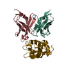
 Assembly


 Mass: 92.094 Da / Num. of mol.: 1 / Source method: obtained synthetically / Formula: C3H8O3
 Mass: 18.015 Da / Num. of mol.: 166 / Source method: isolated from a natural source / Formula: H2O
 Sample preparation
 / Beamline: ID29 / Wavelength: 0.9756
  Processing