 Movie
Movie Controller

[English] 日本語
 Yorodumi
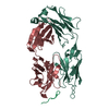
- PDB-4wnd: Crystal structure of the TPR domain of LGN in complex with Frmpd4... -
+ Open data

- Basic information
| Entry | Database: PDB / ID: 4wnd | ||||||
|---|---|---|---|---|---|---|---|
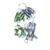
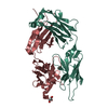
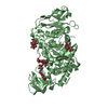
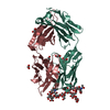
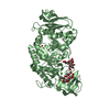
| Title | Crystal structure of the TPR domain of LGN in complex with Frmpd4/Preso1 at 1.5 Angstrom resolution | ||||||
 Components Components |
| ||||||
 Keywords Keywords | SIGNALING PROTEIN/PROTEIN BINDING / TETRATRICOPEPTIDE REPEAT / TPR / CELL POLARITY / CYTOPLASM AND CELL CORTEX / SIGNALING PROTEIN-PROTEIN BINDING COMPLEX | ||||||
| Function / homology |  Function and homology information Function and homology informationmaintenance of centrosome location / positive regulation of synapse structural plasticity / lateral cell cortex / cell cortex region / response to light intensity / positive regulation of spindle assembly / GDP-dissociation inhibitor activity / mitotic spindle pole / neurotransmitter receptor localization to postsynaptic specialization membrane / establishment of mitotic spindle orientation ...maintenance of centrosome location / positive regulation of synapse structural plasticity / lateral cell cortex / cell cortex region / response to light intensity / positive regulation of spindle assembly / GDP-dissociation inhibitor activity / mitotic spindle pole / neurotransmitter receptor localization to postsynaptic specialization membrane / establishment of mitotic spindle orientation / dynein complex binding / lateral plasma membrane / G-protein alpha-subunit binding / phosphatidylinositol-4,5-bisphosphate binding / positive regulation of protein localization to cell cortex / mitotic spindle organization / regulation of mitotic spindle organization / cell cortex / G alpha (i) signalling events / dendritic spine / postsynaptic density / G protein-coupled receptor signaling pathway / protein domain specific binding / cell division / nucleotide binding / centrosome / protein-containing complex / identical protein binding / cytosol / cytoplasm Similarity search - Function | ||||||
| Biological species |  Homo sapiens (human) Homo sapiens (human) | ||||||
| Method |  X-RAY DIFFRACTION / SYNCHROTRON / MOLECULAR REPLACEMENT / Resolution: 1.5 Å X-RAY DIFFRACTION / SYNCHROTRON / MOLECULAR REPLACEMENT / Resolution: 1.5 Å | ||||||
 Authors Authors | Takayanagi, H. / Yuzawa, S. / Sumimoto, H. | ||||||
 Citation Citation | Journal: Acta Crystallogr.,Sect.F / Year: 2015 Title: Structural basis for the recognition of the scaffold protein Frmpd4/Preso1 by the TPR domain of the adaptor protein LGN Authors: Takayanagi, H. / Yuzawa, S. / Sumimoto, H. | ||||||
| History |
|
- Structure visualization
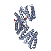
| Structure viewer | Molecule: MolmilJmol/JSmol |
|---|
- Downloads & links
-Download
| PDBx/mmCIF format | 4wnd.cif.gz | 101 KB | Display | PDBx/mmCIF format |
|---|---|---|---|---|
| PDB format | pdb4wnd.ent.gz | 73.1 KB | Display | PDB format |
| PDBx/mmJSON format | 4wnd.json.gz | Tree view | PDBx/mmJSON format | |
| Others |  Other downloads Other downloads |
-Validation report
| Arichive directory | https://data.pdbj.org/pub/pdb/validation_reports/wn/4wndftp://data.pdbj.org/pub/pdb/validation_reports/wn/4wnd | HTTPS FTP |
|---|
-Related structure data
| Related structure data | 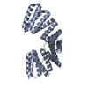 4wneSC  4wnfC 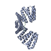 4wngC S: Starting model for refinement C: citing same article ( |
|---|---|
| Similar structure data |


-Links
 PDBj
PDBj


- Assembly
| Deposited unit | 
| ||||||||
|---|---|---|---|---|---|---|---|---|---|
| 1 |
| ||||||||
| Unit cell |
|
-Components
-Protein , 2 types, 2 molecules AB
| #1: Protein | Mass: 44948.281 Da / Num. of mol.: 1 / Fragment: N-terminal TPR domain, UNP residues 20-421 Source method: isolated from a genetically manipulated source Source: (gene. exp.) Homo sapiens (human) / Gene: GPSM2, LGN / Plasmid: pRSFDUET-1 / Production host:  |
|---|---|
| #2: Protein | Mass: 5831.625 Da / Num. of mol.: 1 / Fragment: FRMPD4-L, UNP residues 978-1025 Source method: isolated from a genetically manipulated source Source: (gene. exp.) Homo sapiens (human) / Gene: FRMPD4, KIAA0316, PDZD10, PDZK10 / Plasmid: pGEX-6P-2 / Production host: |
-Non-polymers , 5 types, 317 molecules 


| #3: Chemical | ChemComp-SCN /  Mass: 58.082 Da / Num. of mol.: 7 / Source method: obtained synthetically / Formula: CNS Mass: 58.082 Da / Num. of mol.: 7 / Source method: obtained synthetically / Formula: CNS#4: Chemical | ChemComp-PEG /  Mass: 106.120 Da / Num. of mol.: 5 / Source method: obtained synthetically / Formula: C4H10O3 Mass: 106.120 Da / Num. of mol.: 5 / Source method: obtained synthetically / Formula: C4H10O3#5: Chemical | ChemComp-PGE / |  Mass: 150.173 Da / Num. of mol.: 1 / Source method: obtained synthetically / Formula: C6H14O4 Mass: 150.173 Da / Num. of mol.: 1 / Source method: obtained synthetically / Formula: C6H14O4#6: Chemical | ChemComp-EDO /  Mass: 62.068 Da / Num. of mol.: 6 / Source method: obtained synthetically / Formula: C2H6O2 Mass: 62.068 Da / Num. of mol.: 6 / Source method: obtained synthetically / Formula: C2H6O2#7: Water | ChemComp-HOH / | Mass: 18.015 Da / Num. of mol.: 298 / Source method: isolated from a natural source / Formula: H2O |
|---|
-Experimental details
-Experiment
| Experiment | Method: X-RAY DIFFRACTION / Number of used crystals: 1 |
|---|
- Sample preparation
| Crystal | Density Matthews: 1.96 Å3/Da / Density % sol: 37.22 % / Mosaicity: 0.483 ° |
|---|---|
| Crystal grow | Temperature: 293 K / Method: microbatch / pH: 7.5 Details: 0.1 M BisTrispropane (pH 7.5), 0.2 M potassium thiocyanate, 20% PEG 3350 |
-Data collection
| Diffraction | Mean temperature: 90 K | ||||||||||||||||||||||||||||||||||||||||||||||||||||||||||||||||||||||||||||||||||||||||||||||||||||||||||||||||||||||||||||||||||||||||||||||||||||||||||||||||||||||||||||||||||||||||||||||||||||||||||||||||||
|---|---|---|---|---|---|---|---|---|---|---|---|---|---|---|---|---|---|---|---|---|---|---|---|---|---|---|---|---|---|---|---|---|---|---|---|---|---|---|---|---|---|---|---|---|---|---|---|---|---|---|---|---|---|---|---|---|---|---|---|---|---|---|---|---|---|---|---|---|---|---|---|---|---|---|---|---|---|---|---|---|---|---|---|---|---|---|---|---|---|---|---|---|---|---|---|---|---|---|---|---|---|---|---|---|---|---|---|---|---|---|---|---|---|---|---|---|---|---|---|---|---|---|---|---|---|---|---|---|---|---|---|---|---|---|---|---|---|---|---|---|---|---|---|---|---|---|---|---|---|---|---|---|---|---|---|---|---|---|---|---|---|---|---|---|---|---|---|---|---|---|---|---|---|---|---|---|---|---|---|---|---|---|---|---|---|---|---|---|---|---|---|---|---|---|---|---|---|---|---|---|---|---|---|---|---|---|---|---|---|---|---|
| Diffraction source | Source: SYNCHROTRON / Site: SPring-8  / Beamline: BL44XU / Wavelength: 0.9 Å / Beamline: BL44XU / Wavelength: 0.9 Å | ||||||||||||||||||||||||||||||||||||||||||||||||||||||||||||||||||||||||||||||||||||||||||||||||||||||||||||||||||||||||||||||||||||||||||||||||||||||||||||||||||||||||||||||||||||||||||||||||||||||||||||||||||
| Detector | Type: RAYONIX MX300HE / Detector: CCD / Date: Apr 13, 2014 | ||||||||||||||||||||||||||||||||||||||||||||||||||||||||||||||||||||||||||||||||||||||||||||||||||||||||||||||||||||||||||||||||||||||||||||||||||||||||||||||||||||||||||||||||||||||||||||||||||||||||||||||||||
| Radiation | Protocol: SINGLE WAVELENGTH / Monochromatic (M) / Laue (L): M / Scattering type: x-ray | ||||||||||||||||||||||||||||||||||||||||||||||||||||||||||||||||||||||||||||||||||||||||||||||||||||||||||||||||||||||||||||||||||||||||||||||||||||||||||||||||||||||||||||||||||||||||||||||||||||||||||||||||||
| Radiation wavelength | Wavelength: 0.9 Å / Relative weight: 1 | ||||||||||||||||||||||||||||||||||||||||||||||||||||||||||||||||||||||||||||||||||||||||||||||||||||||||||||||||||||||||||||||||||||||||||||||||||||||||||||||||||||||||||||||||||||||||||||||||||||||||||||||||||
| Reflection | Resolution: 1.5→50 Å / Num. obs: 61319 / % possible obs: 98.8 % / Redundancy: 6.2 % / Rmerge(I) obs: 0.037 / Rpim(I) all: 0.014 / Rrim(I) all: 0.04 / Rsym value: 0.037 / Χ2: 0.986 / Net I/av σ(I): 36.531 / Net I/σ(I): 21.9 / Num. measured all: 379458 | ||||||||||||||||||||||||||||||||||||||||||||||||||||||||||||||||||||||||||||||||||||||||||||||||||||||||||||||||||||||||||||||||||||||||||||||||||||||||||||||||||||||||||||||||||||||||||||||||||||||||||||||||||
| Reflection shell | Diffraction-ID: 1 / Rejects: _
|
- Processing
| Software |
| ||||||||||||||||||||||||||||||||||||||||||||||||||||||||||||
|---|---|---|---|---|---|---|---|---|---|---|---|---|---|---|---|---|---|---|---|---|---|---|---|---|---|---|---|---|---|---|---|---|---|---|---|---|---|---|---|---|---|---|---|---|---|---|---|---|---|---|---|---|---|---|---|---|---|---|---|---|---|
| Refinement | Method to determine structure: MOLECULAR REPLACEMENT Starting model: 4WNE Resolution: 1.5→46.79 Å / Cor.coef. Fo:Fc: 0.973 / Cor.coef. Fo:Fc free: 0.966 / WRfactor Rfree: 0.208 / WRfactor Rwork: 0.1869 / FOM work R set: 0.8847 / SU B: 1.189 / SU ML: 0.044 / SU R Cruickshank DPI: 0.0684 / SU Rfree: 0.0673 / Cross valid method: THROUGHOUT / σ(F): 0 / ESU R: 0.068 / ESU R Free: 0.067 / Stereochemistry target values: MAXIMUM LIKELIHOOD Details: HYDROGENS HAVE BEEN ADDED IN THE RIDING POSITIONS U VALUES : REFINED INDIVIDUALLY
| ||||||||||||||||||||||||||||||||||||||||||||||||||||||||||||
| Solvent computation | Ion probe radii: 0.8 Å / Shrinkage radii: 0.8 Å / VDW probe radii: 1.2 Å / Solvent model: MASK | ||||||||||||||||||||||||||||||||||||||||||||||||||||||||||||
| Displacement parameters | Biso max: 81.59 Å2 / Biso mean: 24.126 Å2 / Biso min: 11.97 Å2
| ||||||||||||||||||||||||||||||||||||||||||||||||||||||||||||
| Refinement step | Cycle: final / Resolution: 1.5→46.79 Å
| ||||||||||||||||||||||||||||||||||||||||||||||||||||||||||||
| Refine LS restraints |
| ||||||||||||||||||||||||||||||||||||||||||||||||||||||||||||
| LS refinement shell | Resolution: 1.504→1.543 Å / Total num. of bins used: 20
|
