[English] 日本語
Yorodumi
- PDB-4wlh: High resolution crystal structure of human kynurenine aminotransf... -

+
Open data


ID or keywords:

Loading...

-
Basic information

Entry
Database: PDB / ID: 4wlh
TitleHigh resolution crystal structure of human kynurenine aminotransferase-I bound to PLP cofactor
ComponentsKynurenine--oxoglutarate transaminase 1
KeywordsTRANSFERASE / Aminotransferase / Alternate conformations / Alpha beta protein
Function / homology
Function and homology information


glutamine-phenylpyruvate transaminase / glutamine-phenylpyruvate transaminase activity / cysteine-S-conjugate beta-lyase activity / L-kynurenine catabolic process / cysteine-S-conjugate beta-lyase / Phenylalanine metabolism / kynurenine-oxoglutarate transaminase / kynurenine-oxoglutarate transaminase activity / kynurenine metabolic process / Glutamate and glutamine metabolism ...glutamine-phenylpyruvate transaminase / glutamine-phenylpyruvate transaminase activity / cysteine-S-conjugate beta-lyase activity / L-kynurenine catabolic process / cysteine-S-conjugate beta-lyase / Phenylalanine metabolism / kynurenine-oxoglutarate transaminase / kynurenine-oxoglutarate transaminase activity / kynurenine metabolic process / Glutamate and glutamine metabolism / Tryptophan catabolism / biosynthetic process / response to bacterium / pyridoxal phosphate binding / protein homodimerization activity / mitochondrion / cytoplasm / cytosol
Similarity search - Function
Aminotransferase, class I/classII / Aminotransferase class I and II / Aspartate Aminotransferase, domain 1 / Aspartate Aminotransferase, domain 1 / Aspartate Aminotransferase; domain 2 / Type I PLP-dependent aspartate aminotransferase-like (Major domain) / Pyridoxal phosphate-dependent transferase, small domain / Pyridoxal phosphate-dependent transferase, major domain / Pyridoxal phosphate-dependent transferase / Alpha-Beta Complex ...Aminotransferase, class I/classII / Aminotransferase class I and II / Aspartate Aminotransferase, domain 1 / Aspartate Aminotransferase, domain 1 / Aspartate Aminotransferase; domain 2 / Type I PLP-dependent aspartate aminotransferase-like (Major domain) / Pyridoxal phosphate-dependent transferase, small domain / Pyridoxal phosphate-dependent transferase, major domain / Pyridoxal phosphate-dependent transferase / Alpha-Beta Complex / 3-Layer(aba) Sandwich / Alpha Beta
Similarity search - Domain/homology
Kynurenine--oxoglutarate transaminase 1
Similarity search - Component
Biological speciesHomo sapiens (human)
MethodX-RAY DIFFRACTION / SYNCHROTRON / MOLECULAR REPLACEMENT / molecular replacement / Resolution: 1.28 Å
Model details
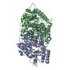
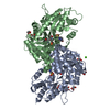
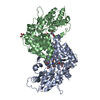
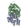
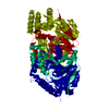
The structure determined at 1.28 Angstroms allowed for the identification of many residues with ...The structure determined at 1.28 Angstroms allowed for the identification of many residues with alternate conformations and the PLP-bound catalytic lysine has a double bond between C4 and NZ which is expected for the internal aldimine form of PLP
AuthorsNadvi, N.A. / Salam, N.K. / Park, J. / Akladios, F.N. / Kapoor, V. / Collyer, C.A. / Gorrell, M.D. / Church, W.B.
CitationJournal: Protein Sci. / Year: 2017
Title: High resolution crystal structures of human kynurenine aminotransferase-I bound to PLP cofactor, and in complex with aminooxyacetate.
Authors: Nadvi, N.A. / Salam, N.K. / Park, J. / Akladios, F.N. / Kapoor, V. / Collyer, C.A. / Gorrell, M.D. / Church, W.B.
History
DepositionOct 7, 2014Deposition site: RCSB / Processing site: RCSB
Revision 1.0Dec 3, 2014Provider: repository / Type: Initial release
Revision 1.1Jul 20, 2016Group: Other
Revision 1.2May 3, 2017Group: Non-polymer description
Revision 1.3May 10, 2017Group: Structure summary
Revision 1.4May 31, 2017Group: Database references
Revision 1.5Sep 27, 2023Group: Advisory / Data collection ...Advisory / Data collection / Database references / Derived calculations / Refinement description
Category: chem_comp_atom / chem_comp_bond ...chem_comp_atom / chem_comp_bond / database_2 / diffrn_radiation_wavelength / pdbx_initial_refinement_model / pdbx_struct_special_symmetry / pdbx_unobs_or_zero_occ_residues
Item: _database_2.pdbx_DOI / _database_2.pdbx_database_accession
Revision 1.6Nov 15, 2023Group: Data collection / Category: chem_comp_atom / chem_comp_bond / Item: _chem_comp_atom.atom_id / _chem_comp_bond.atom_id_2

-
Structure visualization

Structure viewerMolecule:
MolmilJmol/JSmol

Downloads & links

-
Assembly

Deposited unit
A: Kynurenine--oxoglutarate transaminase 1
B: Kynurenine--oxoglutarate transaminase 1


Theoretical massNumber of molelcules
Total (without water)96,3122
Polymers96,3122
Non-polymers00
Water11,313628
1


  • Idetical with deposited unit
  • defined by author&software
TypeNameSymmetry operationNumber
identity operation1_555x,y,z1
Buried area6030 Å2
ΔGint-30 kcal/mol
Surface area31160 Å2
MethodPISA
Unit cell
Length a, b, c (Å)102.440, 107.560, 81.530
Angle α, β, γ (deg.)90.00, 112.64, 90.00
Int Tables number5
Space group name H-MC121
Components on special symmetry positions
IDModelComponents
11A-902-

HOH

Detailsbiological unit is the same as asym.

-
Components

#1: Protein Kynurenine--oxoglutarate transaminase 1 / Cysteine-S-conjugate beta-lyase / Glutamine transaminase K / GTK / Glutamine--phenylpyruvate ...Cysteine-S-conjugate beta-lyase / Glutamine transaminase K / GTK / Glutamine--phenylpyruvate transaminase / Kynurenine aminotransferase 1 / Kynurenine aminotransferase I / KATI / Kynurenine--oxoglutarate transaminase I


Mass: 48156.000 Da / Num. of mol.: 2
Source method: isolated from a genetically manipulated source
Source: (gene. exp.) Homo sapiens (human) / Gene: KYAT1, CCBL1 / Plasmid: PBLUEBAC4.5 / Production host: Spodoptera frugiperda (fall armyworm)
References: UniProt: Q16773, kynurenine-oxoglutarate transaminase, cysteine-S-conjugate beta-lyase, glutamine-phenylpyruvate transaminase
#2: Water ChemComp-HOH / water


Mass: 18.015 Da / Num. of mol.: 628 / Source method: isolated from a natural source / Formula: H2O

-
Experimental details

-
Experiment

ExperimentMethod: X-RAY DIFFRACTION / Number of used crystals: 1

-
Sample preparation

CrystalDensity Matthews: 2.16 Å3/Da / Density % sol: 43.09 %
Crystal growTemperature: 298 K / Method: vapor diffusion, hanging drop / pH: 7.3 / Details: 29% PEG 4000, 0.2 M sodium acetate, 0.1 M Tris

-
Data collection

DiffractionMean temperature: 100 K
Diffraction sourceSource: SYNCHROTRON / Site: Australian Synchrotron / Beamline: MX1 / Wavelength: 0.95369 Å
DetectorType: ADSC QUANTUM 210r / Detector: CCD / Date: Apr 2, 2011
RadiationMonochromator: Silicon Double Crystal / Protocol: SINGLE WAVELENGTH / Monochromatic (M) / Laue (L): M / Scattering type: x-ray
Radiation wavelengthWavelength: 0.95369 Å / Relative weight: 1
ReflectionResolution: 1.28→75.247 Å / Num. obs: 208782 / % possible obs: 100 % / Redundancy: 3.9 % / Biso Wilson estimate: 9.79 Å2 / Rsym value: 0.07 / Net I/σ(I): 11.4
Reflection shellResolution: 1.28→1.35 Å / Redundancy: 3.8 % / Rmerge(I) obs: 0.616 / Mean I/σ(I) obs: 1.3 / Rsym value: 0.616 / % possible all: 100

-
Phasing

PhasingMethod: molecular replacement

-
Processing

Software
NameVersionClassification
PHENIX1.8.4_1496refinement
Blu-Icedata collection
SCALA3.3.16data scaling
PHASER2.1.4phasing
PDB_EXTRACT3.15data extraction
MOSFLMdata reduction
SCALAdata scaling
RefinementMethod to determine structure: MOLECULAR REPLACEMENT
Starting model: 3FVX
Resolution: 1.28→20.98 Å / SU ML: 0.12 / Cross valid method: FREE R-VALUE / σ(F): 1.34 / Phase error: 18.12 / Stereochemistry target values: ML
Details: THE HIGH RESOLUTION AT WHICH THIS STRUCTURE WAS DETERMINED ALLOWED THE IDENTIFICATION OF RESIDUES WITH ALTERNATE CONFORMATIONS. FOUR REGIONS IN EACH CHAIN WERE OBSERVED TO BE HIGHLY ...Details: THE HIGH RESOLUTION AT WHICH THIS STRUCTURE WAS DETERMINED ALLOWED THE IDENTIFICATION OF RESIDUES WITH ALTERNATE CONFORMATIONS. FOUR REGIONS IN EACH CHAIN WERE OBSERVED TO BE HIGHLY DISORDERED WITH GENERALLY POORER ELECTION (419-422). RESIDUES 147-154 WERE REMOVED FROM THE FINAL MODEL DUE TO THE ABSENCE OF UNAMBIGUOUS ELECTION DENSITY. FOR RESIDUES IN THE OTHER DISORDERED REGIONS ELECTRON DENSITY COULD BE OBSERVED FOR MOST OF THE MAIN CHAIN ATOMS, BUT WAS LESS UNAMBIGUOUS FOR THE SIDE-CHAINS. THESE RESIDUES ARE IN THE FINAL MODEL, WITH BOTH THE OCCUPANCY AND TEMPERATURE FACTORS SET TO ZERO FOR MOST OF THESE RESIDUES, DUE TO THE EXISTENCE OF A LEVEL OF EVIDENCE FOR THEIR LOCATION.
RfactorNum. reflection% reflection
Rfree0.187 10493 5.03 %
Rwork0.172 --
obs0.172 208771 100 %
Solvent computationShrinkage radii: 0.9 Å / VDW probe radii: 1.11 Å / Solvent model: FLAT BULK SOLVENT MODEL
Displacement parametersBiso mean: 14.11 Å2
Refinement stepCycle: LAST / Resolution: 1.28→20.98 Å
ProteinNucleic acidLigandSolventTotal
Num. atoms6649 0 0 628 7277
Refine LS restraints
Refine-IDTypeDev idealNumber
X-RAY DIFFRACTIONf_bond_d0.0097301
X-RAY DIFFRACTIONf_angle_d1.3479973
X-RAY DIFFRACTIONf_dihedral_angle_d12.9472730
X-RAY DIFFRACTIONf_chiral_restr0.0781041
X-RAY DIFFRACTIONf_plane_restr0.0081321
LS refinement shell
Resolution (Å)Rfactor RfreeNum. reflection RfreeRfactor RworkNum. reflection RworkRefine-ID% reflection obs (%)
1.28-1.29450.2743470.25936575X-RAY DIFFRACTION100
1.2945-1.30980.29323400.25146587X-RAY DIFFRACTION100
1.3098-1.32570.25323540.23716587X-RAY DIFFRACTION100
1.3257-1.34250.24793300.23436574X-RAY DIFFRACTION100
1.3425-1.36020.25283360.22276605X-RAY DIFFRACTION100
1.3602-1.37880.24553790.21666636X-RAY DIFFRACTION100
1.3788-1.39850.22783260.21226568X-RAY DIFFRACTION100
1.3985-1.41940.22293370.20616611X-RAY DIFFRACTION100
1.4194-1.44160.21753630.20176558X-RAY DIFFRACTION100
1.4416-1.46520.21443570.19186594X-RAY DIFFRACTION100
1.4652-1.49040.22363330.19226608X-RAY DIFFRACTION100
1.4904-1.51750.2083720.17776591X-RAY DIFFRACTION100
1.5175-1.54670.17973600.16956610X-RAY DIFFRACTION100
1.5467-1.57830.18753370.16426604X-RAY DIFFRACTION100
1.5783-1.61260.1753570.15946587X-RAY DIFFRACTION100
1.6126-1.65010.18473640.15816614X-RAY DIFFRACTION100
1.6501-1.69130.1813120.15726643X-RAY DIFFRACTION100
1.6913-1.7370.19323700.15176550X-RAY DIFFRACTION100
1.737-1.78810.17343390.15666610X-RAY DIFFRACTION100
1.7881-1.84580.18983630.16226617X-RAY DIFFRACTION100
1.8458-1.91170.19113310.16876631X-RAY DIFFRACTION100
1.9117-1.98820.17843430.1676603X-RAY DIFFRACTION100
1.9882-2.07860.18663780.16156574X-RAY DIFFRACTION100
2.0786-2.18810.17633450.15996639X-RAY DIFFRACTION100
2.1881-2.3250.14453390.14886654X-RAY DIFFRACTION100
2.325-2.50430.16023460.1526601X-RAY DIFFRACTION100
2.5043-2.75580.16953420.16496672X-RAY DIFFRACTION100
2.7558-3.15340.19613420.17966632X-RAY DIFFRACTION100
3.1534-3.96860.17863760.16846634X-RAY DIFFRACTION100
3.9686-20.97870.16373750.15856709X-RAY DIFFRACTION100

+
About Yorodumi

-
News

-
Feb 9, 2022. New format data for meta-information of EMDB entries

New format data for meta-information of EMDB entries

  • Version 3 of the EMDB header file is now the official format.
  • The previous official version 1.9 will be removed from the archive.

Related info.:EMDB header

External links:wwPDB to switch to version 3 of the EMDB data model

-
Aug 12, 2020. Covid-19 info

Covid-19 info

URL: https://pdbjlvh1.pdbj.org/emnavi/covid19.php

New page: Covid-19 featured information page in EM Navigator.

Related info.:Covid-19 info / Mar 5, 2020. Novel coronavirus structure data

+
Mar 5, 2020. Novel coronavirus structure data

Novel coronavirus structure data

Related info.:Yorodumi Speices / Aug 12, 2020. Covid-19 info

External links:COVID-19 featured content - PDBj / Molecule of the Month (242):Coronavirus Proteases

+
Jan 31, 2019. EMDB accession codes are about to change! (news from PDBe EMDB page)

EMDB accession codes are about to change! (news from PDBe EMDB page)

  • The allocation of 4 digits for EMDB accession codes will soon come to an end. Whilst these codes will remain in use, new EMDB accession codes will include an additional digit and will expand incrementally as the available range of codes is exhausted. The current 4-digit format prefixed with “EMD-” (i.e. EMD-XXXX) will advance to a 5-digit format (i.e. EMD-XXXXX), and so on. It is currently estimated that the 4-digit codes will be depleted around Spring 2019, at which point the 5-digit format will come into force.
  • The EM Navigator/Yorodumi systems omit the EMD- prefix.

Related info.:Q: What is EMD? / ID/Accession-code notation in Yorodumi/EM Navigator

External links:EMDB Accession Codes are Changing Soon! / Contact to PDBj

+
Jul 12, 2017. Major update of PDB

Major update of PDB

  • wwPDB released updated PDB data conforming to the new PDBx/mmCIF dictionary.
  • This is a major update changing the version number from 4 to 5, and with Remediation, in which all the entries are updated.
  • In this update, many items about electron microscopy experimental information are reorganized (e.g. em_software).
  • Now, EM Navigator and Yorodumi are based on the updated data.

External links:wwPDB Remediation / Enriched Model Files Conforming to OneDep Data Standards Now Available in the PDB FTP Archive

-
Yorodumi

Thousand views of thousand structures

  • Yorodumi is a browser for structure data from EMDB, PDB, SASBDB, etc.
  • This page is also the successor to EM Navigator detail page, and also detail information page/front-end page for Omokage search.
  • The word "yorodu" (or yorozu) is an old Japanese word meaning "ten thousand". "mi" (miru) is to see.

Related info.:EMDB / PDB / SASBDB / Comparison of 3 databanks / Yorodumi Search / Aug 31, 2016. New EM Navigator & Yorodumi / Yorodumi Papers / Jmol/JSmol / Function and homology information / Changes in new EM Navigator and Yorodumi

Read more