 Movie
Movie Controller

[English] 日本語
 Yorodumi
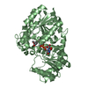
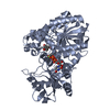
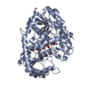
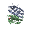
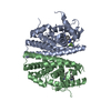
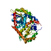
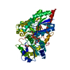
- PDB-4whm: Crystal structure of UDP-glucose: anthocyanidin 3-O-glucosyltrans... -
+ Open data

- Basic information
| Entry | Database: PDB / ID: 4whm | ||||||
|---|---|---|---|---|---|---|---|
| Title | Crystal structure of UDP-glucose: anthocyanidin 3-O-glucosyltransferase in complex with UDP | ||||||
 Components Components | UDP-glucose:anthocyanidin 3-O-glucosyltransferase | ||||||
 Keywords Keywords | TRANSFERASE / Ct3GT-A / UGT78K6 / GT-B fold / Complexed with UDP | ||||||
| Function / homology |  Function and homology information Function and homology informationUDP-glucosyltransferase activity / Transferases; Glycosyltransferases; Hexosyltransferases / nucleotide binding Similarity search - Function | ||||||
| Biological species |  Clitoria ternatea (plant) Clitoria ternatea (plant) | ||||||
| Method |  X-RAY DIFFRACTION / SYNCHROTRON / MOLECULAR REPLACEMENT / Resolution: 1.851 Å X-RAY DIFFRACTION / SYNCHROTRON / MOLECULAR REPLACEMENT / Resolution: 1.851 Å | ||||||
 Authors Authors | Hiromoto, T. / Honjo, E. / Tamada, T. / Kuroki, R. | ||||||
 Citation Citation | Journal: Protein Sci. / Year: 2015 Title: Structural basis for acceptor-substrate recognition of UDP-glucose: anthocyanidin 3-O-glucosyltransferase from Clitoria ternatea Authors: Hiromoto, T. / Honjo, E. / Noda, N. / Tamada, T. / Kazuma, K. / Suzuki, M. / Blaber, M. / Kuroki, R. | ||||||
| History |
|
- Structure visualization
| Structure viewer | Molecule: MolmilJmol/JSmol |
|---|
- Downloads & links
-Download
| PDBx/mmCIF format | 4whm.cif.gz | 197.7 KB | Display | PDBx/mmCIF format |
|---|---|---|---|---|
| PDB format | pdb4whm.ent.gz | 153.4 KB | Display | PDB format |
| PDBx/mmJSON format | 4whm.json.gz | Tree view | PDBx/mmJSON format | |
| Others |  Other downloads Other downloads |
-Validation report
| Arichive directory | https://data.pdbj.org/pub/pdb/validation_reports/wh/4whmftp://data.pdbj.org/pub/pdb/validation_reports/wh/4whm | HTTPS FTP |
|---|
-Related structure data
| Related structure data | 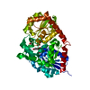 4relC  4remC 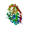 4renC 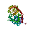 3wc4S S: Starting model for refinement C: citing same article ( |
|---|---|
| Similar structure data |


-Links
 PDBj
PDBj


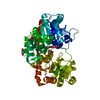
- Assembly
| Deposited unit | 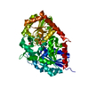
| ||||||||
|---|---|---|---|---|---|---|---|---|---|
| 1 |
| ||||||||
| Unit cell |
|
-Components
| #1: Protein | Mass: 48777.246 Da / Num. of mol.: 1 Source method: isolated from a genetically manipulated source Source: (gene. exp.) Clitoria ternatea (plant) / Gene: Ct3GT-A / Plasmid: pQE-31 / Production host:  References: UniProt: A4F1R4, anthocyanidin 3-O-glucosyltransferase | ||||
|---|---|---|---|---|---|
| #2: Chemical | ChemComp-UDP /   Type: RNA linking / Mass: 404.161 Da / Num. of mol.: 1 / Source method: obtained synthetically / Formula: C9H14N2O12P2 / Comment: UDP*YM Type: RNA linking / Mass: 404.161 Da / Num. of mol.: 1 / Source method: obtained synthetically / Formula: C9H14N2O12P2 / Comment: UDP*YM | ||||
| #3: Chemical |   Mass: 92.094 Da / Num. of mol.: 2 / Source method: obtained synthetically / Formula: C3H8O3 Mass: 92.094 Da / Num. of mol.: 2 / Source method: obtained synthetically / Formula: C3H8O3#4: Chemical | ChemComp-ACT / |   Mass: 59.044 Da / Num. of mol.: 1 / Source method: obtained synthetically / Formula: C2H3O2 Mass: 59.044 Da / Num. of mol.: 1 / Source method: obtained synthetically / Formula: C2H3O2#5: Water | ChemComp-HOH / |  Mass: 18.015 Da / Num. of mol.: 486 / Source method: isolated from a natural source / Formula: H2O Mass: 18.015 Da / Num. of mol.: 486 / Source method: isolated from a natural source / Formula: H2O |
-Experimental details
-Experiment
| Experiment | Method: X-RAY DIFFRACTION / Number of used crystals: 1 |
|---|
- Sample preparation
| Crystal | Density Matthews: 2.35 Å3/Da / Density % sol: 47.55 % |
|---|---|
| Crystal grow | Temperature: 293 K / Method: vapor diffusion, hanging drop / pH: 5.6 Details: 0.2M ammonium acetate, 26%(w/v) PEG 4000, 0.1M sodium citrate |
-Data collection
| Diffraction | Mean temperature: 100 K | |||||||||||||||||||||||||||||||||||||||||||||||||||||||||||||||||||||||||||||
|---|---|---|---|---|---|---|---|---|---|---|---|---|---|---|---|---|---|---|---|---|---|---|---|---|---|---|---|---|---|---|---|---|---|---|---|---|---|---|---|---|---|---|---|---|---|---|---|---|---|---|---|---|---|---|---|---|---|---|---|---|---|---|---|---|---|---|---|---|---|---|---|---|---|---|---|---|---|---|
| Diffraction source | Source: SYNCHROTRON / Site: SPring-8  / Beamline: BL38B1 / Wavelength: 1 Å / Beamline: BL38B1 / Wavelength: 1 Å | |||||||||||||||||||||||||||||||||||||||||||||||||||||||||||||||||||||||||||||
| Detector | Type: ADSC QUANTUM 315r / Detector: CCD / Date: Jul 5, 2008 | |||||||||||||||||||||||||||||||||||||||||||||||||||||||||||||||||||||||||||||
| Radiation | Monochromator: Si(111) double crystal / Protocol: SINGLE WAVELENGTH / Monochromatic (M) / Laue (L): M / Scattering type: x-ray | |||||||||||||||||||||||||||||||||||||||||||||||||||||||||||||||||||||||||||||
| Radiation wavelength | Wavelength: 1 Å / Relative weight: 1 | |||||||||||||||||||||||||||||||||||||||||||||||||||||||||||||||||||||||||||||
| Reflection | Resolution: 1.85→50 Å / Num. obs: 38569 / % possible obs: 99.6 % / Redundancy: 3.7 % / Biso Wilson estimate: 16.45 Å2 / Rmerge(I) obs: 0.083 / Χ2: 0.915 / Net I/av σ(I): 15.2 / Net I/σ(I): 8.8 / Num. measured all: 142408 | |||||||||||||||||||||||||||||||||||||||||||||||||||||||||||||||||||||||||||||
| Reflection shell | Diffraction-ID: 1 / Rejects: _
|
- Processing
| Software |
| |||||||||||||||||||||||||||||||||||||||||||||||||||||||||||||||||||||||||||||||||||||||||||||||||||||||||
|---|---|---|---|---|---|---|---|---|---|---|---|---|---|---|---|---|---|---|---|---|---|---|---|---|---|---|---|---|---|---|---|---|---|---|---|---|---|---|---|---|---|---|---|---|---|---|---|---|---|---|---|---|---|---|---|---|---|---|---|---|---|---|---|---|---|---|---|---|---|---|---|---|---|---|---|---|---|---|---|---|---|---|---|---|---|---|---|---|---|---|---|---|---|---|---|---|---|---|---|---|---|---|---|---|---|---|
| Refinement | Method to determine structure: MOLECULAR REPLACEMENT Starting model: 3WC4 Resolution: 1.851→26.235 Å / FOM work R set: 0.8926 / SU ML: 0.18 / Cross valid method: THROUGHOUT / σ(F): 1.36 / Phase error: 17.99 / Stereochemistry target values: ML
| |||||||||||||||||||||||||||||||||||||||||||||||||||||||||||||||||||||||||||||||||||||||||||||||||||||||||
| Solvent computation | Shrinkage radii: 0.9 Å / VDW probe radii: 1.11 Å / Solvent model: FLAT BULK SOLVENT MODEL | |||||||||||||||||||||||||||||||||||||||||||||||||||||||||||||||||||||||||||||||||||||||||||||||||||||||||
| Displacement parameters | Biso max: 72.71 Å2 / Biso mean: 16.48 Å2 / Biso min: 4.24 Å2 | |||||||||||||||||||||||||||||||||||||||||||||||||||||||||||||||||||||||||||||||||||||||||||||||||||||||||
| Refinement step | Cycle: final / Resolution: 1.851→26.235 Å
| |||||||||||||||||||||||||||||||||||||||||||||||||||||||||||||||||||||||||||||||||||||||||||||||||||||||||
| Refine LS restraints |
| |||||||||||||||||||||||||||||||||||||||||||||||||||||||||||||||||||||||||||||||||||||||||||||||||||||||||
| LS refinement shell |
| |||||||||||||||||||||||||||||||||||||||||||||||||||||||||||||||||||||||||||||||||||||||||||||||||||||||||
| Refinement TLS params. | Method: refined / Refine-ID: X-RAY DIFFRACTION
| |||||||||||||||||||||||||||||||||||||||||||||||||||||||||||||||||||||||||||||||||||||||||||||||||||||||||
| Refinement TLS group |
|
