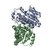
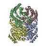
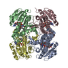
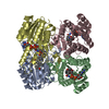
[English] 日本語
Yorodumi
- PDB-4w7i: Crystal structure of DEH reductase A1-R' mutant -

+
Open data


ID or keywords:

Loading...

-
Basic information

Entry
Database: PDB / ID: 4w7i
TitleCrystal structure of DEH reductase A1-R' mutant
Components4-deoxy-L-erythro-5-hexoseulose uronate reductase A1-R'
KeywordsOXIDOREDUCTASE / ALPHA/BETA/ALPHA / ROSSMANN-FOLD / Alginate metabolism / Short-chain dehydrogenase/reductase / loop-exchange
Function / homologyEnoyl-(Acyl carrier protein) reductase / Short-chain dehydrogenase/reductase SDR / NAD(P)-binding Rossmann-like Domain / NAD(P)-binding domain superfamily / Rossmann fold / 3-Layer(aba) Sandwich / Alpha Beta / 4-deoxy-L-erythro-5-hexoseulose uronate reductase A1-R'
Function and homology information
Biological speciesSphingomonas sp. A1 (bacteria)
MethodX-RAY DIFFRACTION / SYNCHROTRON / MOLECULAR REPLACEMENT / Resolution: 1.98 Å
AuthorsTakase, R. / Mikami, B. / Kawai, S. / Murata, K. / Hashimoto, W.
Funding support Japan, 4items
OrganizationGrant numberCountry
Grants-in-Aid for Scientific Research from Japan Society for the Promotion of Science Japan
Promotion of Basic Research Activities for Innovative Biosciences (PROBRAIN) of Japan Japan
Targeted Proteins Research Program from the Ministry of Education, Culture, Sports, Science, and Technology (MEXT) of Japan Japan
Research fellowships from Japan Society for the Promotion of Science for Young Scientists Japan
CitationJournal: J.Biol.Chem. / Year: 2014
Title: Structure-based Conversion of the Coenzyme Requirement of a Short-chain Dehydrogenase/Reductase Involved in Bacterial Alginate Metabolism.
Authors: Takase, R. / Mikami, B. / Kawai, S. / Murata, K. / Hashimoto, W.
History
DepositionAug 22, 2014Deposition site: RCSB / Processing site: PDBJ
Revision 1.0Sep 24, 2014Provider: repository / Type: Initial release
Revision 1.1Dec 10, 2014Group: Database references
Revision 1.2Jan 29, 2020Group: Data collection / Database references ...Data collection / Database references / Derived calculations / Other / Source and taxonomy
Category: citation / diffrn_source ...citation / diffrn_source / entity_src_gen / pdbx_database_status / pdbx_prerelease_seq / pdbx_struct_oper_list
Item: _citation.journal_id_CSD / _diffrn_source.pdbx_synchrotron_site ..._citation.journal_id_CSD / _diffrn_source.pdbx_synchrotron_site / _entity_src_gen.pdbx_alt_source_flag / _pdbx_database_status.pdb_format_compatible / _pdbx_struct_oper_list.symmetry_operation
Revision 1.3Nov 8, 2023Group: Data collection / Database references / Refinement description
Category: chem_comp_atom / chem_comp_bond ...chem_comp_atom / chem_comp_bond / database_2 / pdbx_initial_refinement_model / refine_hist
Item: _database_2.pdbx_DOI / _database_2.pdbx_database_accession ..._database_2.pdbx_DOI / _database_2.pdbx_database_accession / _refine_hist.number_atoms_solvent / _refine_hist.pdbx_number_atoms_ligand / _refine_hist.pdbx_number_atoms_nucleic_acid / _refine_hist.pdbx_number_atoms_protein

-
Structure visualization

Structure viewerMolecule:
MolmilJmol/JSmol

Downloads & links

-
Assembly

Deposited unit
A: 4-deoxy-L-erythro-5-hexoseulose uronate reductase A1-R'
B: 4-deoxy-L-erythro-5-hexoseulose uronate reductase A1-R'
C: 4-deoxy-L-erythro-5-hexoseulose uronate reductase A1-R'
D: 4-deoxy-L-erythro-5-hexoseulose uronate reductase A1-R'


Theoretical massNumber of molelcules
Total (without water)109,4134
Polymers109,4134
Non-polymers00
Water4,035224
1
A: 4-deoxy-L-erythro-5-hexoseulose uronate reductase A1-R'
B: 4-deoxy-L-erythro-5-hexoseulose uronate reductase A1-R'

A: 4-deoxy-L-erythro-5-hexoseulose uronate reductase A1-R'
B: 4-deoxy-L-erythro-5-hexoseulose uronate reductase A1-R'


Theoretical massNumber of molelcules
Total (without water)109,4134
Polymers109,4134
Non-polymers00
Water724
TypeNameSymmetry operationNumber
identity operation1_555x,y,z1
crystal symmetry operation2_454-x-1,y,-z-11
Buried area12810 Å2
ΔGint-69 kcal/mol
Surface area33370 Å2
MethodPISA
2
C: 4-deoxy-L-erythro-5-hexoseulose uronate reductase A1-R'
D: 4-deoxy-L-erythro-5-hexoseulose uronate reductase A1-R'

C: 4-deoxy-L-erythro-5-hexoseulose uronate reductase A1-R'
D: 4-deoxy-L-erythro-5-hexoseulose uronate reductase A1-R'


Theoretical massNumber of molelcules
Total (without water)109,4134
Polymers109,4134
Non-polymers00
Water724
TypeNameSymmetry operationNumber
identity operation1_555x,y,z1
crystal symmetry operation2_555-x,y,-z1
Buried area13070 Å2
ΔGint-73 kcal/mol
Surface area32910 Å2
MethodPISA
Unit cell
Length a, b, c (Å)71.215, 178.152, 96.013
Angle α, β, γ (deg.)90.00, 111.76, 90.00
Int Tables number5
Space group name H-MC121
Components on special symmetry positions
IDModelComponents
11A-311-

HOH

21B-302-

HOH

31B-317-

HOH

-
Components

#1: Protein
4-deoxy-L-erythro-5-hexoseulose uronate reductase A1-R'


Mass: 27353.174 Da / Num. of mol.: 4 / Mutation: T16S, E17Q, N37H, S38G, H39R, V40K, D41A
Source method: isolated from a genetically manipulated source
Source: (gene. exp.) Sphingomonas sp. A1 (bacteria) / Production host: Escherichia coli BL21(DE3) (bacteria) / References: UniProt: A0A096LNF1*PLUS
#2: Water ChemComp-HOH / water


Mass: 18.015 Da / Num. of mol.: 224 / Source method: isolated from a natural source / Formula: H2O
Sequence detailsTHIS PROTEIN IS MUTANT FORM WITH MUTATION SITES T16S, E17Q, N37H, S38G, H39R, V40K, AND D41A. ...THIS PROTEIN IS MUTANT FORM WITH MUTATION SITES T16S, E17Q, N37H, S38G, H39R, V40K, AND D41A. SEQUENCE OF THE WILD TYPE HAS BEEN DEPOSITED TO GENBANK WITH ACCESSION ID AB970509.

-
Experimental details

-
Experiment

ExperimentMethod: X-RAY DIFFRACTION / Number of used crystals: 1

-
Sample preparation

CrystalDensity Matthews: 2.59 Å3/Da / Density % sol: 52.42 %
Crystal growTemperature: 293 K / Method: vapor diffusion / pH: 6.5 / Details: PEG 300, cacodylate, calcium acetate

-
Data collection

DiffractionMean temperature: 100 K
Diffraction sourceSource: SYNCHROTRON / Site: SPring-8 / Beamline: BL38B1 / Wavelength: 1 Å
DetectorType: ADSC QUANTUM 315 / Detector: CCD / Date: Jul 18, 2014
RadiationProtocol: SINGLE WAVELENGTH / Monochromatic (M) / Laue (L): M / Scattering type: x-ray
Radiation wavelengthWavelength: 1 Å / Relative weight: 1
ReflectionResolution: 1.98→50 Å / Num. obs: 77352 / % possible obs: 99.8 % / Redundancy: 4.2 % / Net I/σ(I): 35.9
Reflection shellResolution: 1.98→2.01 Å / Redundancy: 4.2 % / % possible all: 100

-
Processing

SoftwareName: REFMAC / Version: 5.8.0049 / Classification: refinement
RefinementMethod to determine structure: MOLECULAR REPLACEMENT
Starting model: 4TKL
Resolution: 1.98→50 Å / Cor.coef. Fo:Fc: 0.947 / Cor.coef. Fo:Fc free: 0.918 / SU B: 6.242 / SU ML: 0.169 / Cross valid method: THROUGHOUT / ESU R: 0.199 / ESU R Free: 0.185 / Stereochemistry target values: MAXIMUM LIKELIHOOD / Details: HYDROGENS HAVE BEEN ADDED IN THE RIDING POSITIONS
RfactorNum. reflection% reflectionSelection details
Rfree0.28618 3882 5 %RANDOM
Rwork0.23688 ---
obs0.23932 73314 99.66 %-
Solvent computationIon probe radii: 0.8 Å / Shrinkage radii: 0.8 Å / VDW probe radii: 1.2 Å / Solvent model: MASK
Displacement parametersBiso mean: 41.11 Å2
Baniso -1Baniso -2Baniso -3
1--1.47 Å2-0 Å20.23 Å2
2--0.27 Å2-0 Å2
3---0.77 Å2
Refinement stepCycle: 1 / Resolution: 1.98→50 Å
ProteinNucleic acidLigandSolventTotal
Num. atoms7320 0 0 224 7544
Refine LS restraints
Refine-IDTypeDev idealDev ideal targetNumber
X-RAY DIFFRACTIONr_bond_refined_d0.0120.0197444
X-RAY DIFFRACTIONr_bond_other_d0.0010.027253
X-RAY DIFFRACTIONr_angle_refined_deg1.4541.94910042
X-RAY DIFFRACTIONr_angle_other_deg0.825316577
X-RAY DIFFRACTIONr_dihedral_angle_1_deg6.3835977
X-RAY DIFFRACTIONr_dihedral_angle_2_deg40.85922.632304
X-RAY DIFFRACTIONr_dihedral_angle_3_deg15.235151211
X-RAY DIFFRACTIONr_dihedral_angle_4_deg16.5651564
X-RAY DIFFRACTIONr_chiral_restr0.0790.21129
X-RAY DIFFRACTIONr_gen_planes_refined0.0070.028571
X-RAY DIFFRACTIONr_gen_planes_other0.0010.021765
X-RAY DIFFRACTIONr_nbd_refined
X-RAY DIFFRACTIONr_nbd_other
X-RAY DIFFRACTIONr_nbtor_refined
X-RAY DIFFRACTIONr_nbtor_other
X-RAY DIFFRACTIONr_xyhbond_nbd_refined
X-RAY DIFFRACTIONr_xyhbond_nbd_other
X-RAY DIFFRACTIONr_metal_ion_refined
X-RAY DIFFRACTIONr_metal_ion_other
X-RAY DIFFRACTIONr_symmetry_vdw_refined
X-RAY DIFFRACTIONr_symmetry_vdw_other
X-RAY DIFFRACTIONr_symmetry_hbond_refined
X-RAY DIFFRACTIONr_symmetry_hbond_other
X-RAY DIFFRACTIONr_symmetry_metal_ion_refined
X-RAY DIFFRACTIONr_symmetry_metal_ion_other
X-RAY DIFFRACTIONr_mcbond_it2.7673.9023932
X-RAY DIFFRACTIONr_mcbond_other2.7653.9023931
X-RAY DIFFRACTIONr_mcangle_it4.0145.8434901
X-RAY DIFFRACTIONr_mcangle_other4.0135.8434902
X-RAY DIFFRACTIONr_scbond_it2.8414.2873512
X-RAY DIFFRACTIONr_scbond_other2.8414.2873513
X-RAY DIFFRACTIONr_scangle_it
X-RAY DIFFRACTIONr_scangle_other4.2636.2795142
X-RAY DIFFRACTIONr_long_range_B_refined6.32532.1088790
X-RAY DIFFRACTIONr_long_range_B_other6.31532.0848751
X-RAY DIFFRACTIONr_rigid_bond_restr
X-RAY DIFFRACTIONr_sphericity_free
X-RAY DIFFRACTIONr_sphericity_bonded
LS refinement shellResolution: 1.976→2.027 Å / Total num. of bins used: 20
RfactorNum. reflection% reflection
Rfree0.431 279 -
Rwork0.369 5326 -
obs--98.39 %

+
About Yorodumi

-
News

-
Feb 9, 2022. New format data for meta-information of EMDB entries

New format data for meta-information of EMDB entries

  • Version 3 of the EMDB header file is now the official format.
  • The previous official version 1.9 will be removed from the archive.

Related info.:EMDB header

External links:wwPDB to switch to version 3 of the EMDB data model

-
Aug 12, 2020. Covid-19 info

Covid-19 info

URL: https://pdbjlvh1.pdbj.org/emnavi/covid19.php

New page: Covid-19 featured information page in EM Navigator.

Related info.:Covid-19 info / Mar 5, 2020. Novel coronavirus structure data

+
Mar 5, 2020. Novel coronavirus structure data

Novel coronavirus structure data

Related info.:Yorodumi Speices / Aug 12, 2020. Covid-19 info

External links:COVID-19 featured content - PDBj / Molecule of the Month (242):Coronavirus Proteases

+
Jan 31, 2019. EMDB accession codes are about to change! (news from PDBe EMDB page)

EMDB accession codes are about to change! (news from PDBe EMDB page)

  • The allocation of 4 digits for EMDB accession codes will soon come to an end. Whilst these codes will remain in use, new EMDB accession codes will include an additional digit and will expand incrementally as the available range of codes is exhausted. The current 4-digit format prefixed with “EMD-” (i.e. EMD-XXXX) will advance to a 5-digit format (i.e. EMD-XXXXX), and so on. It is currently estimated that the 4-digit codes will be depleted around Spring 2019, at which point the 5-digit format will come into force.
  • The EM Navigator/Yorodumi systems omit the EMD- prefix.

Related info.:Q: What is EMD? / ID/Accession-code notation in Yorodumi/EM Navigator

External links:EMDB Accession Codes are Changing Soon! / Contact to PDBj

+
Jul 12, 2017. Major update of PDB

Major update of PDB

  • wwPDB released updated PDB data conforming to the new PDBx/mmCIF dictionary.
  • This is a major update changing the version number from 4 to 5, and with Remediation, in which all the entries are updated.
  • In this update, many items about electron microscopy experimental information are reorganized (e.g. em_software).
  • Now, EM Navigator and Yorodumi are based on the updated data.

External links:wwPDB Remediation / Enriched Model Files Conforming to OneDep Data Standards Now Available in the PDB FTP Archive

-
Yorodumi

Thousand views of thousand structures

  • Yorodumi is a browser for structure data from EMDB, PDB, SASBDB, etc.
  • This page is also the successor to EM Navigator detail page, and also detail information page/front-end page for Omokage search.
  • The word "yorodu" (or yorozu) is an old Japanese word meaning "ten thousand". "mi" (miru) is to see.

Related info.:EMDB / PDB / SASBDB / Comparison of 3 databanks / Yorodumi Search / Aug 31, 2016. New EM Navigator & Yorodumi / Yorodumi Papers / Jmol/JSmol / Function and homology information / Changes in new EM Navigator and Yorodumi

Read more