 Movie
Movie Controller

[English] 日本語
 Yorodumi
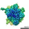
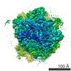
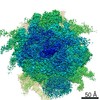
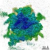
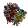
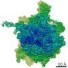
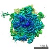
- PDB-4v6i: Localization of the small subunit ribosomal proteins into a 6.1 A... -
+ Open data

- Basic information
| Entry | Database: PDB / ID: 4v6i | |||||||||
|---|---|---|---|---|---|---|---|---|---|---|
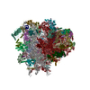
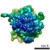
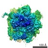
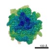
| Title | Localization of the small subunit ribosomal proteins into a 6.1 A cryo-EM map of Saccharomyces cerevisiae translating 80S ribosome | |||||||||
 Components Components |
| |||||||||
 Keywords Keywords | RIBOSOME / eukaryotic ribosome / homology modeling / de novo modeling / ribosomal proteins / novel ribosomal proteins | |||||||||
| Function / homology |  Function and homology information Function and homology information: / cytoplasmic translational elongation / regulation of amino acid metabolic process / negative regulation of glucose mediated signaling pathway / positive regulation of translational fidelity / RMTs methylate histone arginines / Protein methylation / Protein hydroxylation / ribosome-associated ubiquitin-dependent protein catabolic process / GDP-dissociation inhibitor activity ...: / cytoplasmic translational elongation / regulation of amino acid metabolic process / negative regulation of glucose mediated signaling pathway / positive regulation of translational fidelity / RMTs methylate histone arginines / Protein methylation / Protein hydroxylation / ribosome-associated ubiquitin-dependent protein catabolic process / GDP-dissociation inhibitor activity / positive regulation of nuclear-transcribed mRNA catabolic process, deadenylation-dependent decay / pre-mRNA 5'-splice site binding / Formation of the ternary complex, and subsequently, the 43S complex / Translation initiation complex formation / Ribosomal scanning and start codon recognition / preribosome, small subunit precursor / nonfunctional rRNA decay / response to cycloheximide / cleavage in ITS2 between 5.8S rRNA and LSU-rRNA of tricistronic rRNA transcript (SSU-rRNA, 5.8S rRNA, LSU-rRNA) / Major pathway of rRNA processing in the nucleolus and cytosol / mRNA destabilization / SRP-dependent cotranslational protein targeting to membrane / GTP hydrolysis and joining of the 60S ribosomal subunit / Nonsense Mediated Decay (NMD) independent of the Exon Junction Complex (EJC) / Nonsense Mediated Decay (NMD) enhanced by the Exon Junction Complex (EJC) / negative regulation of translational frameshifting / Formation of a pool of free 40S subunits / negative regulation of mRNA splicing, via spliceosome / preribosome, large subunit precursor / L13a-mediated translational silencing of Ceruloplasmin expression / endonucleolytic cleavage to generate mature 3'-end of SSU-rRNA from (SSU-rRNA, 5.8S rRNA, LSU-rRNA) / ribosomal large subunit export from nucleus / translational elongation / G-protein alpha-subunit binding / protein kinase activator activity / 90S preribosome / ribosomal subunit export from nucleus / regulation of translational fidelity / ribonucleoprotein complex binding / endonucleolytic cleavage in ITS1 to separate SSU-rRNA from 5.8S rRNA and LSU-rRNA from tricistronic rRNA transcript (SSU-rRNA, 5.8S rRNA, LSU-rRNA) / protein-RNA complex assembly / translational termination / maturation of LSU-rRNA / ribosomal small subunit export from nucleus / translation regulator activity / DNA-(apurinic or apyrimidinic site) endonuclease activity / rescue of stalled ribosome / protein kinase C binding / maturation of LSU-rRNA from tricistronic rRNA transcript (SSU-rRNA, 5.8S rRNA, LSU-rRNA) / ribosomal large subunit biogenesis / maturation of SSU-rRNA from tricistronic rRNA transcript (SSU-rRNA, 5.8S rRNA, LSU-rRNA) / maturation of SSU-rRNA / small-subunit processome / translational initiation / macroautophagy / maintenance of translational fidelity / modification-dependent protein catabolic process / protein tag activity / cytoplasmic stress granule / rRNA processing / ribosome biogenesis / large ribosomal subunit / ribosome binding / ribosomal small subunit biogenesis / ribosomal small subunit assembly / small ribosomal subunit / small ribosomal subunit rRNA binding / 5S rRNA binding / ribosomal large subunit assembly / cytosolic small ribosomal subunit / large ribosomal subunit rRNA binding / cytosolic large ribosomal subunit / cytoplasmic translation / negative regulation of translation / rRNA binding / protein ubiquitination / structural constituent of ribosome / ribosome / translation / G protein-coupled receptor signaling pathway / negative regulation of gene expression / response to antibiotic / mRNA binding / ubiquitin protein ligase binding / nucleolus / mitochondrion / RNA binding / zinc ion binding / nucleoplasm / metal ion binding / nucleus / cytoplasm / cytosol Similarity search - Function | |||||||||
| Biological species |  | |||||||||
| Method | ELECTRON MICROSCOPY / single particle reconstruction / cryo EM / Resolution: 8.8 Å | |||||||||
 Authors Authors | Armache, J.-P. / Jarasch, A. / Anger, A.M. / Villa, E. / Becker, T. / Bhushan, S. / Jossinet, F. / Habeck, M. / Dindar, G. / Franckenberg, S. ...Armache, J.-P. / Jarasch, A. / Anger, A.M. / Villa, E. / Becker, T. / Bhushan, S. / Jossinet, F. / Habeck, M. / Dindar, G. / Franckenberg, S. / Marquez, V. / Mielke, T. / Thomm, M. / Berninghausen, O. / Beatrix, B. / Soeding, J. / Westhof, E. / Wilson, D.N. / Beckmann, R. | |||||||||
 Citation Citation | Journal: Proc Natl Acad Sci U S A / Year: 2010 Title: Cryo-EM structure and rRNA model of a translating eukaryotic 80S ribosome at 5.5-A resolution. Authors: Jean-Paul Armache / Alexander Jarasch / Andreas M Anger / Elizabeth Villa / Thomas Becker / Shashi Bhushan / Fabrice Jossinet / Michael Habeck / Gülcin Dindar / Sibylle Franckenberg / Viter ...Authors: Jean-Paul Armache / Alexander Jarasch / Andreas M Anger / Elizabeth Villa / Thomas Becker / Shashi Bhushan / Fabrice Jossinet / Michael Habeck / Gülcin Dindar / Sibylle Franckenberg / Viter Marquez / Thorsten Mielke / Michael Thomm / Otto Berninghausen / Birgitta Beatrix / Johannes Söding / Eric Westhof / Daniel N Wilson / Roland Beckmann /  Abstract: Protein biosynthesis, the translation of the genetic code into polypeptides, occurs on ribonucleoprotein particles called ribosomes. Although X-ray structures of bacterial ribosomes are available, ...Protein biosynthesis, the translation of the genetic code into polypeptides, occurs on ribonucleoprotein particles called ribosomes. Although X-ray structures of bacterial ribosomes are available, high-resolution structures of eukaryotic 80S ribosomes are lacking. Using cryoelectron microscopy and single-particle reconstruction, we have determined the structure of a translating plant (Triticum aestivum) 80S ribosome at 5.5-Å resolution. This map, together with a 6.1-Å map of a Saccharomyces cerevisiae 80S ribosome, has enabled us to model ∼98% of the rRNA. Accurate assignment of the rRNA expansion segments (ES) and variable regions has revealed unique ES-ES and r-protein-ES interactions, providing insight into the structure and evolution of the eukaryotic ribosome. #1: Journal: Proc Natl Acad Sci U S A / Year: 2010Title: Localization of eukaryote-specific ribosomal proteins in a 5.5-Å cryo-EM map of the 80S eukaryotic ribosome. Authors: Jean-Paul Armache / Alexander Jarasch / Andreas M Anger / Elizabeth Villa / Thomas Becker / Shashi Bhushan / Fabrice Jossinet / Michael Habeck / Gülcin Dindar / Sibylle Franckenberg / Viter ...Authors: Jean-Paul Armache / Alexander Jarasch / Andreas M Anger / Elizabeth Villa / Thomas Becker / Shashi Bhushan / Fabrice Jossinet / Michael Habeck / Gülcin Dindar / Sibylle Franckenberg / Viter Marquez / Thorsten Mielke / Michael Thomm / Otto Berninghausen / Birgitta Beatrix / Johannes Söding / Eric Westhof / Daniel N Wilson / Roland Beckmann / Abstract: Protein synthesis in all living organisms occurs on ribonucleoprotein particles, called ribosomes. Despite the universality of this process, eukaryotic ribosomes are significantly larger in size than ...Protein synthesis in all living organisms occurs on ribonucleoprotein particles, called ribosomes. Despite the universality of this process, eukaryotic ribosomes are significantly larger in size than their bacterial counterparts due in part to the presence of 80 r proteins rather than 54 in bacteria. Using cryoelectron microscopy reconstructions of a translating plant (Triticum aestivum) 80S ribosome at 5.5-Å resolution, together with a 6.1-Å map of a translating Saccharomyces cerevisiae 80S ribosome, we have localized and modeled 74/80 (92.5%) of the ribosomal proteins, encompassing 12 archaeal/eukaryote-specific small subunit proteins as well as the complete complement of the ribosomal proteins of the eukaryotic large subunit. Near-complete atomic models of the 80S ribosome provide insights into the structure, function, and evolution of the eukaryotic translational apparatus. | |||||||||
| History |
|
- Structure visualization
| Movie |
Movie viewer |
|---|---|
| Structure viewer | Molecule: MolmilJmol/JSmol |
- Downloads & links
-Download
| PDBx/mmCIF format | 4v6i.cif.gz | 4.5 MB | Display | PDBx/mmCIF format |
|---|---|---|---|---|
| PDB format | pdb4v6i.ent.gz | Display | PDB format | |
| PDBx/mmJSON format | 4v6i.json.gz | Tree view | PDBx/mmJSON format | |
| Others |  Other downloads Other downloads |
-Validation report
| Summary document | 4v6i_validation.pdf.gz | 1.9 MB | Display | wwPDB validaton report |
|---|---|---|---|---|
| Full document | 4v6i_full_validation.pdf.gz | 4.5 MB | Display | |
| Data in XML | 4v6i_validation.xml.gz | 563.1 KB | Display | |
| Data in CIF | 4v6i_validation.cif.gz | 860.6 KB | Display | |
| Arichive directory | https://data.pdbj.org/pub/pdb/validation_reports/v6/4v6iftp://data.pdbj.org/pub/pdb/validation_reports/v6/4v6i | HTTPS FTP |
-Related structure data
| Related structure data | 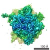 1669M  3izdC  3iz5 3iz6 3iz7 3iz9 C: citing same article ( M: map data used to model this data |
|---|---|
| Similar structure data |

-Links
 PDBj
PDBj


- Assembly
| Deposited unit | 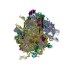
|
|---|---|
| 1 |
|
-Components
+40S ribosomal protein ... , 27 types, 27 molecules AaAAABADACAEAGAFAHAIAJAKALAMANAOAQAPARASATAVAWAXAYAZAU
-Unknown 40S ribosomal protein ... , 2 types, 2 molecules AbAc
| #27: Protein/peptide | Mass: 3166.895 Da / Num. of mol.: 1 / Source method: isolated from a natural source / Source: (natural) |
|---|---|
| #28: Protein/peptide | Mass: 2230.741 Da / Num. of mol.: 1 / Source method: isolated from a natural source / Source: (natural) |
+60S ribosomal protein ... , 43 types, 43 molecules BABBBCBDBEBGBFBHBJBKBNBMBPBOBRBTBUBWBVBXBZBYBaBdBcBfBeBgBhBi...
-60S acidic ribosomal protein ... , 3 types, 5 molecules BsBtBuBvBw
| #38: Protein | Mass: 33749.121 Da / Num. of mol.: 1 / Source method: isolated from a natural source / Source: (natural) | ||
|---|---|---|---|
| #72: Protein | Mass: 10928.809 Da / Num. of mol.: 2 / Source method: isolated from a natural source / Source: (natural) #73: Protein | Mass: 10752.715 Da / Num. of mol.: 2 / Source method: isolated from a natural source / Source: (natural) |
-Protein/peptide , 2 types, 3 molecules BxByBz
| #70: Protein/peptide | Mass: 1805.216 Da / Num. of mol.: 2 / Source method: isolated from a natural source / Source: (natural) #71: Protein/peptide | | Mass: 1294.587 Da / Num. of mol.: 1 / Source method: isolated from a natural source / Source: (natural) |
|---|
-RNA chain , 6 types, 6 molecules CACBCCDADBDC
| #78: RNA chain | Mass: 579761.938 Da / Num. of mol.: 1 / Source method: isolated from a natural source / Source: (natural) |
|---|---|
| #79: RNA chain | Mass: 24135.262 Da / Num. of mol.: 1 / Source method: isolated from a natural source / Source: (natural) |
| #80: RNA chain | Mass: 3498.178 Da / Num. of mol.: 1 / Source method: isolated from a natural source / Source: (natural) |
| #81: RNA chain | Mass: 1097493.875 Da / Num. of mol.: 1 / Source method: isolated from a natural source / Source: (natural) |
| #82: RNA chain | Mass: 50682.922 Da / Num. of mol.: 1 / Source method: isolated from a natural source / Source: (natural) |
| #83: RNA chain | Mass: 37947.492 Da / Num. of mol.: 1 / Source method: isolated from a natural source / Source: (natural) |
-Experimental details
-Experiment
| Experiment | Method: ELECTRON MICROSCOPY |
|---|---|
| EM experiment | Aggregation state: PARTICLE / 3D reconstruction method: single particle reconstruction |
- Sample preparation
| Component | Name: Saccharomyces cerevisiae translating 80S ribosome / Type: RIBOSOME |
|---|---|
| Buffer solution | pH: 7.5 |
| Specimen | Embedding applied: NO / Shadowing applied: NO / Staining applied: NO / Vitrification applied: YES |
| Vitrification | Instrument: FEI VITROBOT MARK I / Cryogen name: ETHANE |
- Electron microscopy imaging
| Experimental equipment |  Model: Tecnai Polara / Image courtesy: FEI Company |
|---|---|
| Microscopy | Model: FEI POLARA 300 |
| Electron gun | Electron source:  FIELD EMISSION GUN / Accelerating voltage: 300 kV / Illumination mode: FLOOD BEAM FIELD EMISSION GUN / Accelerating voltage: 300 kV / Illumination mode: FLOOD BEAM |
| Electron lens | Mode: BRIGHT FIELD / Nominal magnification: 39000 X / Nominal defocus max: 4500 nm / Nominal defocus min: 1200 nm |
| Image recording | Electron dose: 25 e/Å2 / Film or detector model: KODAK SO-163 FILM |
| Radiation | Protocol: SINGLE WAVELENGTH / Monochromatic (M) / Laue (L): M / Scattering type: x-ray |
| Radiation wavelength | Relative weight: 1 |
- Processing
| Symmetry | Point symmetry: C1 (asymmetric) | ||||||||||||
|---|---|---|---|---|---|---|---|---|---|---|---|---|---|
| 3D reconstruction | Resolution: 8.8 Å / Num. of particles: 20400 / Symmetry type: POINT | ||||||||||||
| Refinement step | Cycle: LAST
|
