 Movie
Movie Controller

[English] 日本語
 Yorodumi
- PDB-4v3p: The molecular structure of the left-handed supra-molecular helix ... -
+ Open data

- Basic information
| Entry | Database: PDB / ID: 4v3p | ||||||
|---|---|---|---|---|---|---|---|
| Title | The molecular structure of the left-handed supra-molecular helix of eukaryotic polyribosomes | ||||||
 Components Components |
| ||||||
 Keywords Keywords | RIBOSOME | ||||||
| Function / homology |  Function and homology information Function and homology informationcytoplasmic translational elongation / translational elongation / protein kinase activator activity / ribonucleoprotein complex binding / cytosolic ribosome / maturation of LSU-rRNA from tricistronic rRNA transcript (SSU-rRNA, 5.8S rRNA, LSU-rRNA) / small ribosomal subunit / large ribosomal subunit rRNA binding / cytosolic small ribosomal subunit / cytosolic large ribosomal subunit ...cytoplasmic translational elongation / translational elongation / protein kinase activator activity / ribonucleoprotein complex binding / cytosolic ribosome / maturation of LSU-rRNA from tricistronic rRNA transcript (SSU-rRNA, 5.8S rRNA, LSU-rRNA) / small ribosomal subunit / large ribosomal subunit rRNA binding / cytosolic small ribosomal subunit / cytosolic large ribosomal subunit / cytoplasmic translation / negative regulation of translation / structural constituent of ribosome / ribosome / translation / ribonucleoprotein complex / mRNA binding / signal transduction / RNA binding / zinc ion binding / plasma membrane Similarity search - Function | ||||||
| Biological species |  | ||||||
| Method | ELECTRON MICROSCOPY / subtomogram averaging / cryo EM / Resolution: 34 Å | ||||||
 Authors Authors | Myasnikov, A.G. / Afonina, Z.A. / Menetret, J.F. / Shirokov, V.A. / Spirin, A.S. / Klaholz, B.P. | ||||||
 Citation Citation | Journal: Nat Commun / Year: 2014 Title: The molecular structure of the left-handed supra-molecular helix of eukaryotic polyribosomes. Authors: Alexander G Myasnikov / Zhanna A Afonina / Jean-François Ménétret / Vladimir A Shirokov / Alexander S Spirin / Bruno P Klaholz /   Abstract: During protein synthesis, several ribosomes bind to a single messenger RNA (mRNA) forming large macromolecular assemblies called polyribosomes. Here we report the detailed molecular structure of a ...During protein synthesis, several ribosomes bind to a single messenger RNA (mRNA) forming large macromolecular assemblies called polyribosomes. Here we report the detailed molecular structure of a 100 MDa eukaryotic poly-ribosome complex derived from cryo electron tomography, sub-tomogram averaging and pseudo-atomic modelling by crystal structure fitting. The structure allowed the visualization of the three functional parts of the polysome assembly, the central core region that forms a rather compact left-handed supra-molecular helix, and the more open regions that harbour the initiation and termination sites at either ends. The helical region forms a continuous mRNA channel where the mRNA strand bridges neighbouring exit and entry sites of the ribosomes and prevents mRNA looping between ribosomes. This structure provides unprecedented insights into protein- and RNA-mediated inter-ribosome contacts that involve conserved sites through 40S subunits and long protruding RNA expansion segments, suggesting a role in stabilizing the overall polyribosomal assembly. | ||||||
| History |
|
- Structure visualization
| Movie |
Movie viewer |
|---|---|
| Structure viewer | Molecule: MolmilJmol/JSmol |
- Downloads & links
-Download
| PDBx/mmCIF format | 4v3p.cif.gz | 4.6 MB | Display | PDBx/mmCIF format |
|---|---|---|---|---|
| PDB format | pdb4v3p.ent.gz | Display | PDB format | |
| PDBx/mmJSON format | 4v3p.json.gz | Tree view | PDBx/mmJSON format | |
| Others |  Other downloads Other downloads |
-Validation report
| Arichive directory | https://data.pdbj.org/pub/pdb/validation_reports/v3/4v3pftp://data.pdbj.org/pub/pdb/validation_reports/v3/4v3p | HTTPS FTP |
|---|
-Related structure data
| Related structure data |  2790MC M: map data used to model this data C: citing same article ( |
|---|---|
| Similar structure data |
-Links
 PDBj
PDBj


- Assembly
| Deposited unit | 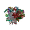
|
|---|---|
| 1 | x 23
|
-Components
+Protein , 5 types, 5 molecules SaSBLALMLh
+40S ribosomal protein ... , 21 types, 21 molecules SASDSESFSISJSKSLSMSOSQSPSSSRSVSYSZSXSCSNST
+40S WHEAT GERM ... , 2 types, 2 molecules SWS3
+Unknown 40S wheat germ ribosome protein ... , 4 types, 4 molecules ScSbSUSG
+Ribosomal protein ... , 12 types, 13 molecules SHLELPLTLlLoLiLjLtLuLeLCLK
+RNA chain , 5 types, 5 molecules S2S1L1L3L2
+60S ribosomal protein ... , 27 types, 27 molecules LBLFLHLOLRLQLULVLXLZLYLbLdLfLgLnLrLGLSLWLaLkLJLcLDLmLI
+Unknown 60S wheat germ ribosome protein ... , 5 types, 6 molecules LqLyLxLzLLLN
+Protein/peptide , 1 types, 1 molecules Lp
+60S acidic ribosomal protein ... , 2 types, 3 molecules LvLwLs
+Details
-Experimental details
-Experiment
| Experiment | Method: ELECTRON MICROSCOPY |
|---|---|
| EM experiment | Aggregation state: PARTICLE / 3D reconstruction method: subtomogram averaging |
- Sample preparation
| Component | Name: POLY-RIBOSOME FROM IN VITRO WHEAT GERM SYSTEM / Type: RIBOSOME |
|---|---|
| Buffer solution | Name: 25MM HEPES-KOH, 3MM MG(OAC)2, 85MM KOAC,1.6MM DTT, 0.25MM SPERMIDINE pH: 7.6 Details: 25MM HEPES-KOH, 3MM MG(OAC)2, 85MM KOAC,1.6MM DTT, 0.25MM SPERMIDINE |
| Specimen | Embedding applied: NO / Shadowing applied: NO / Staining applied: NO / Vitrification applied: YES |
| Specimen support | Details: HOLEY CARBON |
| Vitrification | Instrument: FEI VITROBOT MARK IV / Cryogen name: ETHANE Details: VITRIFICATION 1 --CRYOGEN- ETHANE, HUMIDITY-95, TEMPERATURE- 120,INSTRUMENT- FEI VITROBOT MARK IV, METHOD- 3UL OF SAMPLE APPLIED ON 300 MESH HOLY CARBON QUANTIFOIL GRID. BLOTTING WAS DONE IN ...Details: VITRIFICATION 1 --CRYOGEN- ETHANE, HUMIDITY-95, TEMPERATURE- 120,INSTRUMENT- FEI VITROBOT MARK IV, METHOD- 3UL OF SAMPLE APPLIED ON 300 MESH HOLY CARBON QUANTIFOIL GRID. BLOTTING WAS DONE IN VITROBOT MARK IV, BLOT TIME 0.5 SEC, BLOT FORCE 5 |
- Electron microscopy imaging
| Experimental equipment |  Model: Tecnai F30 / Image courtesy: FEI Company |
|---|---|
| Microscopy | Model: FEI TECNAI F30 / Date: Jan 1, 2013 |
| Electron gun | Electron source:  FIELD EMISSION GUN / Accelerating voltage: 150 kV / Illumination mode: FLOOD BEAM FIELD EMISSION GUN / Accelerating voltage: 150 kV / Illumination mode: FLOOD BEAM |
| Electron lens | Mode: BRIGHT FIELD / Nominal magnification: 39000 X / Calibrated magnification: 41176 X / Nominal defocus max: 4000 nm / Nominal defocus min: 2000 nm / Cs: 2 mm |
| Specimen holder | Temperature: 90 K / Tilt angle max: 70 ° / Tilt angle min: -70 ° |
| Image recording | Electron dose: 30 e/Å2 / Film or detector model: FEI FALCON I (4k x 4k) |
- Processing
| EM software |
| |||||||||||||||
|---|---|---|---|---|---|---|---|---|---|---|---|---|---|---|---|---|
| 3D reconstruction | Method: MAXIMUM LIKELIHOOD / Resolution: 34 Å / Num. of particles: 106 / Actual pixel size: 6.8 Å / Details: SUBMISSION BASED ON EXPERIMENTAL DATA / Symmetry type: HELICAL | |||||||||||||||
| Atomic model building | Protocol: RIGID BODY FIT / Space: REAL / Details: METHOD--RIGID BODY REFINEMENT PROTOCOL--X-RAY | |||||||||||||||
| Atomic model building | PDB-ID: 3IZ6 3iz6 Accession code: 3IZ6 / Source name: PDB / Type: experimental model | |||||||||||||||
| Refinement | Highest resolution: 34 Å |
