 Movie
Movie Controller

[English] 日本語
 Yorodumi
- PDB-3izq: Structure of the Dom34-Hbs1-GDPNP complex bound to a translating ... -
+ Open data

- Basic information
| Entry | Database: PDB / ID: 3izq | ||||||
|---|---|---|---|---|---|---|---|
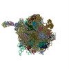
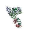
| Title | Structure of the Dom34-Hbs1-GDPNP complex bound to a translating ribosome | ||||||
 Components Components |
| ||||||
 Keywords Keywords | RIBOSOMAL PROTEIN / HYDROLASE / No-Go mRNA decay | ||||||
| Function / homology |  Function and homology information Function and homology informationEukaryotic Translation Elongation / Dom34-Hbs1 complex / RNA surveillance / nuclear-transcribed mRNA catabolic process, no-go decay / nuclear-transcribed mRNA catabolic process, non-stop decay / HSF1 activation / Protein methylation / ribosome disassembly / nonfunctional rRNA decay / positive regulation of translational initiation ...Eukaryotic Translation Elongation / Dom34-Hbs1 complex / RNA surveillance / nuclear-transcribed mRNA catabolic process, no-go decay / nuclear-transcribed mRNA catabolic process, non-stop decay / HSF1 activation / Protein methylation / ribosome disassembly / nonfunctional rRNA decay / positive regulation of translational initiation / translation elongation factor activity / Neutrophil degranulation / RNA endonuclease activity / rescue of stalled cytosolic ribosome / meiotic cell cycle / positive regulation of translation / Hydrolases; Acting on acid anhydrides; Acting on GTP to facilitate cellular and subcellular movement / translation / cell division / GTPase activity / GTP binding / metal ion binding / cytoplasm / cytosol Similarity search - Function | ||||||
| Biological species |  | ||||||
| Method | ELECTRON MICROSCOPY / single particle reconstruction / cryo EM / Resolution: 9.5 Å | ||||||
 Authors Authors | Becker, T. / Armache, J.-P. / Jarasch, A. / Anger, A.M. / Villa, E. / Sieber, H. / Abdel Motaal, B. / Mielke, T. / Berninghausen, O. / Beckmann, R. | ||||||
 Citation Citation | Journal: Nat Struct Mol Biol / Year: 2011 Title: Structure of the no-go mRNA decay complex Dom34-Hbs1 bound to a stalled 80S ribosome. Authors: Thomas Becker / Jean-Paul Armache / Alexander Jarasch / Andreas M Anger / Elizabeth Villa / Heidemarie Sieber / Basma Abdel Motaal / Thorsten Mielke / Otto Berninghausen / Roland Beckmann /  Abstract: No-go decay (NGD) is a mRNA quality-control mechanism in eukaryotic cells that leads to degradation of mRNAs stalled during translational elongation. The key factors triggering NGD are Dom34 and Hbs1. ...No-go decay (NGD) is a mRNA quality-control mechanism in eukaryotic cells that leads to degradation of mRNAs stalled during translational elongation. The key factors triggering NGD are Dom34 and Hbs1. We used cryo-EM to visualize NGD intermediates resulting from binding of the Dom34-Hbs1 complex to stalled ribosomes. At subnanometer resolution, all domains of Dom34 and Hbs1 were identified, allowing the docking of crystal structures and homology models. Moreover, the close structural similarity of Dom34 and Hbs1 to eukaryotic release factors (eRFs) enabled us to propose a model for the ribosome-bound eRF1-eRF3 complex. Collectively, our data provide structural insights into how stalled mRNA is recognized on the ribosome and how the eRF complex can simultaneously recognize stop codons and catalyze peptide release. | ||||||
| History |
|
- Structure visualization
| Movie |
Movie viewer |
|---|---|
| Structure viewer | Molecule: MolmilJmol/JSmol |
- Downloads & links
-Download
| PDBx/mmCIF format | 3izq.cif.gz | 174.9 KB | Display | PDBx/mmCIF format |
|---|---|---|---|---|
| PDB format | pdb3izq.ent.gz | 129.1 KB | Display | PDB format |
| PDBx/mmJSON format | 3izq.json.gz | Tree view | PDBx/mmJSON format | |
| Others |  Other downloads Other downloads |
-Validation report
| Arichive directory | https://data.pdbj.org/pub/pdb/validation_reports/iz/3izqftp://data.pdbj.org/pub/pdb/validation_reports/iz/3izq | HTTPS FTP |
|---|
-Related structure data
| Related structure data | 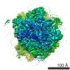 1811MC 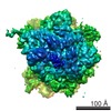 1808C  1809C 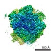 1812C  3izc M: map data used to model this data C: citing same article ( |
|---|---|
| Similar structure data |


-Links
 PDBj
PDBj


- Assembly
| Deposited unit | 
|
|---|---|
| 1 |
|
-Components
| #1: Protein | Mass: 44119.797 Da / Num. of mol.: 1 Source method: isolated from a genetically manipulated source Source: (gene. exp.) Gene: DOM34, N2016, YNL001W / Production host:  References: UniProt: P33309, Hydrolases; Acting on ester bonds |
|---|---|
| #2: Protein | Mass: 68826.406 Da / Num. of mol.: 1 Source method: isolated from a genetically manipulated source Source: (gene. exp.) Gene: HBS1, YKR084C, YKR404 / Production host: |
-Experimental details
-Experiment
| Experiment | Method: ELECTRON MICROSCOPY |
|---|---|
| EM experiment | Aggregation state: PARTICLE / 3D reconstruction method: single particle reconstruction |
- Sample preparation
| Component |
| ||||||||||||
|---|---|---|---|---|---|---|---|---|---|---|---|---|---|
| Buffer solution | pH: 7 | ||||||||||||
| Specimen | Conc.: 0.02 mg/ml / Embedding applied: NO / Shadowing applied: NO / Staining applied: NO / Vitrification applied: YES | ||||||||||||
| Specimen support | Details: Carbon-coated Quantifoil with 2 nm carbon on top | ||||||||||||
| Vitrification | Instrument: FEI VITROBOT MARK I / Cryogen name: ETHANE / Humidity: 100 % Details: Blotted for 10 seconds before plunging, used 2 layer of filter paper |
- Electron microscopy imaging
| Experimental equipment |  Model: Tecnai Polara / Image courtesy: FEI Company |
|---|---|
| Microscopy | Model: FEI POLARA 300 |
| Electron gun | Electron source:  FIELD EMISSION GUN / Accelerating voltage: 300 kV / Illumination mode: FLOOD BEAM FIELD EMISSION GUN / Accelerating voltage: 300 kV / Illumination mode: FLOOD BEAM |
| Electron lens | Mode: BRIGHT FIELD / Nominal magnification: 39000 X / Calibrated magnification: 39000 X / Nominal defocus max: 4000 nm / Nominal defocus min: 1300 nm / Cs: 2.26 mm |
| Specimen holder | Temperature: 84 K |
| Image recording | Electron dose: 25 e/Å2 / Film or detector model: KODAK SO-163 FILM |
| Image scans | Num. digital images: 41 |
| Radiation | Protocol: SINGLE WAVELENGTH / Monochromatic (M) / Laue (L): M / Scattering type: x-ray |
| Radiation wavelength | Relative weight: 1 |
- Processing
| EM software |
| ||||||||||||
|---|---|---|---|---|---|---|---|---|---|---|---|---|---|
| Symmetry | Point symmetry: C1 (asymmetric) | ||||||||||||
| 3D reconstruction | Method: Single particle projection-matching / Resolution: 9.5 Å / Num. of particles: 38400 / Nominal pixel size: 1.2375 Å / Actual pixel size: 1.2375 Å / Symmetry type: POINT | ||||||||||||
| Atomic model building | Space: REAL | ||||||||||||
| Refinement step | Cycle: LAST
|
