 Movie
Movie Controller

[English] 日本語
 Yorodumi
- PDB-4v27: Structure of the GH99 endo-alpha-mannanase from Bacteroides xylan... -
+ Open data

- Basic information
| Entry | Database: PDB / ID: 4v27 | ||||||
|---|---|---|---|---|---|---|---|
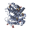
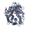
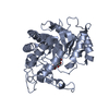
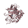
| Title | Structure of the GH99 endo-alpha-mannanase from Bacteroides xylanisolvens in complex with mannose-alpha-1,3-isofagomine | ||||||
 Components Components | GLYCOSYL HYDROLASE FAMILY 71 | ||||||
 Keywords Keywords | HYDROLASE / GH99 / CAZY / MANNAN / BACTEROIDES / POLYSACCHARIDE UTILISATION / ENZYME-CARBOHYDRATE INTERACTION / INHIBITOR / GLYCOSIDASE INHIBITION | ||||||
| Function / homology |  Function and homology information Function and homology information | ||||||
| Biological species |  BACTEROIDES XYLANISOLVENS (bacteria) BACTEROIDES XYLANISOLVENS (bacteria) | ||||||
| Method |  X-RAY DIFFRACTION / MOLECULAR REPLACEMENT / Resolution: 1.9 Å X-RAY DIFFRACTION / MOLECULAR REPLACEMENT / Resolution: 1.9 Å | ||||||
 Authors Authors | Hakki, Z. / Bellmaine, S. / Thompson, A.J. / Speciale, G. / Davies, G.J. / Williams, S.J. | ||||||
 Citation Citation | Journal: Chemistry / Year: 2015 Title: Structural and Kinetic Dissection of the Endo-Alpha-1,2-Mannanase Activity of Bacterial Gh99 Glycoside Hydrolases from Bacteroides Spp. Authors: Hakki, Z. / Thompson, A.J. / Bellmaine, S. / Speciale, G. / Davies, G.J. / Williams, S.J. | ||||||
| History |
|
- Structure visualization
| Structure viewer | Molecule: MolmilJmol/JSmol |
|---|
- Downloads & links
-Download
| PDBx/mmCIF format | 4v27.cif.gz | 93.5 KB | Display | PDBx/mmCIF format |
|---|---|---|---|---|
| PDB format | pdb4v27.ent.gz | 70.4 KB | Display | PDB format |
| PDBx/mmJSON format | 4v27.json.gz | Tree view | PDBx/mmJSON format | |
| Others |  Other downloads Other downloads |
-Validation report
| Arichive directory | https://data.pdbj.org/pub/pdb/validation_reports/v2/4v27ftp://data.pdbj.org/pub/pdb/validation_reports/v2/4v27 | HTTPS FTP |
|---|
-Related structure data
| Related structure data | 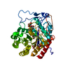 4v28C  4ad1S C: citing same article ( S: Starting model for refinement |
|---|---|
| Similar structure data |


-Links
 PDBj
PDBj


- Assembly
| Deposited unit | 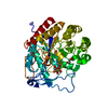
| ||||||||
|---|---|---|---|---|---|---|---|---|---|
| 1 |
| ||||||||
| Unit cell |
| ||||||||
| Components on special symmetry positions |
|
-Components
| #1: Protein | Mass: 43479.266 Da / Num. of mol.: 1 Source method: isolated from a genetically manipulated source Source: (gene. exp.) BACTEROIDES XYLANISOLVENS (bacteria) / Strain: XB1A / Plasmid: PET28A / Production host: References: UniProt: D6D1V7, glycoprotein endo-alpha-1,2-mannosidase | ||
|---|---|---|---|
| #2: Sugar | ChemComp-MAN /   Type: D-saccharide, alpha linking / Mass: 180.156 Da / Num. of mol.: 1 Type: D-saccharide, alpha linking / Mass: 180.156 Da / Num. of mol.: 1Source method: isolated from a genetically manipulated source Formula: C6H12O6 | ||
| #3: Chemical | ChemComp-IFM /   Mass: 147.172 Da / Num. of mol.: 1 / Source method: obtained synthetically / Formula: C6H13NO3 Mass: 147.172 Da / Num. of mol.: 1 / Source method: obtained synthetically / Formula: C6H13NO3 | ||
| #4: Chemical |   Mass: 62.068 Da / Num. of mol.: 3 / Source method: obtained synthetically / Formula: C2H6O2 Mass: 62.068 Da / Num. of mol.: 3 / Source method: obtained synthetically / Formula: C2H6O2#5: Water | ChemComp-HOH / |  Mass: 18.015 Da / Num. of mol.: 430 / Source method: isolated from a natural source / Formula: H2O Mass: 18.015 Da / Num. of mol.: 430 / Source method: isolated from a natural source / Formula: H2O |
-Experimental details
-Experiment
| Experiment | Method: X-RAY DIFFRACTION / Number of used crystals: 1 |
|---|
- Sample preparation
| Crystal | Density Matthews: 2.38 Å3/Da / Density % sol: 48.3 % / Description: NONE |
|---|---|
| Crystal grow | pH: 6.4 / Details: 2.8 M SODIUM ACETATE PH 6.4 |
-Data collection
| Diffraction | Mean temperature: 120 K |
|---|---|
| Diffraction source | Source: ROTATING ANODE / Type: RIGAKU RUH3R / Wavelength: 1.5418 |
| Detector | Type: MAR scanner 345 mm plate / Detector: IMAGE PLATE / Date: Jun 29, 2011 |
| Radiation | Protocol: SINGLE WAVELENGTH / Monochromatic (M) / Laue (L): M / Scattering type: x-ray |
| Radiation wavelength | Wavelength: 1.5418 Å / Relative weight: 1 |
| Reflection | Resolution: 1.9→39.4 Å / Num. obs: 30894 / % possible obs: 100 % / Observed criterion σ(I): -3.7 / Redundancy: 6.1 % / Rmerge(I) obs: 0.05 / Net I/σ(I): 23.2 |
| Reflection shell | Resolution: 1.9→2 Å / Redundancy: 5.9 % / Rmerge(I) obs: 0.2 / Mean I/σ(I) obs: 8.2 / % possible all: 100 |
- Processing
| Software |
| ||||||||||||||||||||||||||||||||||||||||||||||||||||||||||||||||||||||||||||||||||||||||||||||||||||||||||||||||||||||||||||||||||||||||||||||||||||||||||||||||||||||||||||||||||||||
|---|---|---|---|---|---|---|---|---|---|---|---|---|---|---|---|---|---|---|---|---|---|---|---|---|---|---|---|---|---|---|---|---|---|---|---|---|---|---|---|---|---|---|---|---|---|---|---|---|---|---|---|---|---|---|---|---|---|---|---|---|---|---|---|---|---|---|---|---|---|---|---|---|---|---|---|---|---|---|---|---|---|---|---|---|---|---|---|---|---|---|---|---|---|---|---|---|---|---|---|---|---|---|---|---|---|---|---|---|---|---|---|---|---|---|---|---|---|---|---|---|---|---|---|---|---|---|---|---|---|---|---|---|---|---|---|---|---|---|---|---|---|---|---|---|---|---|---|---|---|---|---|---|---|---|---|---|---|---|---|---|---|---|---|---|---|---|---|---|---|---|---|---|---|---|---|---|---|---|---|---|---|---|---|
| Refinement | Method to determine structure: MOLECULAR REPLACEMENT Starting model: PDB ENTRY 4AD1 Resolution: 1.9→38.28 Å / Cor.coef. Fo:Fc: 0.961 / Cor.coef. Fo:Fc free: 0.937 / SU B: 2.361 / SU ML: 0.071 / Cross valid method: THROUGHOUT / ESU R: 0.132 / ESU R Free: 0.122 / Stereochemistry target values: MAXIMUM LIKELIHOOD Details: HYDROGENS HAVE BEEN ADDED IN THE RIDING POSITIONS. HYDROGENS HAVE BEEN USED IF PRESENT IN THE INPUT. U VALUES REFINED INDIVIDUALLY
| ||||||||||||||||||||||||||||||||||||||||||||||||||||||||||||||||||||||||||||||||||||||||||||||||||||||||||||||||||||||||||||||||||||||||||||||||||||||||||||||||||||||||||||||||||||||
| Solvent computation | Ion probe radii: 0.8 Å / Shrinkage radii: 0.8 Å / VDW probe radii: 1.2 Å / Solvent model: MASK | ||||||||||||||||||||||||||||||||||||||||||||||||||||||||||||||||||||||||||||||||||||||||||||||||||||||||||||||||||||||||||||||||||||||||||||||||||||||||||||||||||||||||||||||||||||||
| Displacement parameters | Biso mean: 17.173 Å2
| ||||||||||||||||||||||||||||||||||||||||||||||||||||||||||||||||||||||||||||||||||||||||||||||||||||||||||||||||||||||||||||||||||||||||||||||||||||||||||||||||||||||||||||||||||||||
| Refinement step | Cycle: LAST / Resolution: 1.9→38.28 Å
| ||||||||||||||||||||||||||||||||||||||||||||||||||||||||||||||||||||||||||||||||||||||||||||||||||||||||||||||||||||||||||||||||||||||||||||||||||||||||||||||||||||||||||||||||||||||
| Refine LS restraints |
|