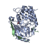
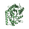
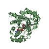
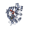
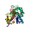
Mass: 33921.035 Da / Num. of mol.: 1 / Fragment: RESIDUES 7-300 Source method: isolated from a genetically manipulated source Details: TRUNCATED MOUSE PP1G INCLUDING RESIDUES OF 7-300 / Source: (gene. exp.) MUS MUSCULUS (house mouse) / Production host: ESCHERICHIA COLI BL21 (bacteria) References: UniProt: P63087, protein-serine/threonine phosphatase
#2: Protein
PROTEINPHOSPHATASE1REGULATORYSUBUNIT15B
Mass: 6927.684 Da / Num. of mol.: 1 / Fragment: RESIDUES 631-684 Source method: isolated from a genetically manipulated source Details: TRUNCATED HUMAN PPP1R15B INCLUDING RESIDUES OF 631-684 Source: (gene. exp.) HOMO SAPIENS (human) / Production host: ESCHERICHIA COLI (E. coli) / References: UniProt: Q5SWA1
Protocol: SINGLE WAVELENGTH / Monochromatic (M) / Laue (L): M / Scattering type: x-ray
Radiation wavelength
Wavelength: 0.979 Å / Relative weight: 1
Reflection
Resolution: 1.85→51.34 Å / Num. obs: 32078 / % possible obs: 100 % / Observed criterion σ(I): 1.5 / Redundancy: 7.8 % / Biso Wilson estimate: 28.03 Å2 / Rmerge(I) obs: 0.09 / Net I/σ(I): 9.7
Reflection shell
Resolution: 1.85→1.89 Å / Redundancy: 8.2 % / Rmerge(I) obs: 0.98 / Mean I/σ(I) obs: 1.5 / % possible all: 100
-
Processing
Software
Name
Version
Classification
PHENIX
(PHENIX.REFINE)
refinement
MOSFLM
datareduction
Aimless
datascaling
CCP4I
phasing
Refinement
Method to determine structure: MOLECULAR REPLACEMENT Starting model: THE CRYSTAL STRUCTURE OF MOUSE PP1G IN COMPLEX WITH TRUNCATED HUMAN PPP1R15B INCLUDING 631-660 Resolution: 1.85→51.337 Å / SU ML: 0.22 / σ(F): 1.34 / Phase error: 23.64 / Stereochemistry target values: ML
Rfactor
Num. reflection
% reflection
Rfree
0.2222
1626
5.1 %
Rwork
0.1766
-
-
obs
0.1789
32078
99.83 %
Solvent computation
Shrinkage radii: 0.9 Å / VDW probe radii: 1.11 Å / Solvent model: FLAT BULK SOLVENT MODEL
Displacement parameters
Biso mean: 45.24 Å2
Refinement step
Cycle: LAST / Resolution: 1.85→51.337 Å
Protein
Nucleic acid
Ligand
Solvent
Total
Num. atoms
2568
0
1
88
2657
Refine LS restraints
Refine-ID
Type
Dev ideal
Number
X-RAY DIFFRACTION
f_bond_d
0.011
2625
X-RAY DIFFRACTION
f_angle_d
1.221
3542
X-RAY DIFFRACTION
f_dihedral_angle_d
14.737
982
X-RAY DIFFRACTION
f_chiral_restr
0.054
378
X-RAY DIFFRACTION
f_plane_restr
0.006
460
LS refinement shell
Resolution (Å)
Rfactor Rfree
Num. reflection Rfree
Rfactor Rwork
Num. reflection Rwork
Refine-ID
% reflection obs (%)
1.85-1.9044
0.368
144
0.3002
2484
X-RAY DIFFRACTION
99
1.9044-1.9659
0.3144
130
0.2567
2458
X-RAY DIFFRACTION
100
1.9659-2.0362
0.2819
130
0.2243
2498
X-RAY DIFFRACTION
100
2.0362-2.1177
0.2668
122
0.2282
2517
X-RAY DIFFRACTION
100
2.1177-2.2141
0.2441
122
0.1989
2499
X-RAY DIFFRACTION
100
2.2141-2.3308
0.2335
142
0.19
2507
X-RAY DIFFRACTION
100
2.3308-2.4769
0.2239
141
0.1892
2494
X-RAY DIFFRACTION
100
2.4769-2.6681
0.2169
133
0.179
2532
X-RAY DIFFRACTION
100
2.6681-2.9366
0.2232
149
0.1833
2523
X-RAY DIFFRACTION
100
2.9366-3.3614
0.2053
141
0.169
2561
X-RAY DIFFRACTION
100
3.3614-4.2347
0.1831
134
0.1463
2598
X-RAY DIFFRACTION
100
4.2347-51.3572
0.2139
138
0.157
2781
X-RAY DIFFRACTION
100
Refinement TLS params.
Method: refined / Refine-ID: X-RAY DIFFRACTION
ID
L11 (°2)
L12 (°2)
L13 (°2)
L22 (°2)
L23 (°2)
L33 (°2)
S11 (Å °)
S12 (Å °)
S13 (Å °)
S21 (Å °)
S22 (Å °)
S23 (Å °)
S31 (Å °)
S32 (Å °)
S33 (Å °)
T11 (Å2)
T12 (Å2)
T13 (Å2)
T22 (Å2)
T23 (Å2)
T33 (Å2)
Origin x (Å)
Origin y (Å)
Origin z (Å)
1
4.5024
2.9413
-0.218
8.0689
-1.309
0.8358
0.2739
0.4815
0.7297
-0.3726
0.3588
0.1215
-0.9545
0.8092
-0.4545
0.6075
-0.2187
0.2441
0.7301
-0.0142
0.4373
-23.4704
21.6744
-25.8765
2
8.4463
4.7294
-4.1416
2.9322
-2.2561
4.9547
0.0157
1.0148
0.3776
-0.7073
0.5721
0.6508
-0.1017
-1.0263
-0.6093
0.2735
-0.0344
-0.0111
0.5544
0.0769
0.2708
-37.2734
14.8031
-21.1595
3
2.555
0.3667
-0.4669
2.1858
0.5333
1.9775
-0.0255
-0.1018
0.3347
-0.1246
0.4415
-0.3529
0.0034
0.7664
-0.2288
0.2128
-0.0468
0.0544
0.5348
-0.1086
0.3068
-22.5376
13.4724
-11.0586
4
1.9728
-1.1596
-1.3771
2.2652
1.1618
4.0105
-0.0065
0.0308
0.2045
-0.0713
0.3237
0.0219
-0.0923
-0.1156
-0.3128
0.1245
-0.0583
0.0166
0.3205
0.0324
0.2453
-33.1692
16.0312
-9.7302
5
5.9506
-0.455
1.6612
4.9215
0.9653
8.1055
-0.1303
0.1806
0.0576
0.1721
0.188
0.7572
0.5231
-0.8757
-0.048
0.2168
-0.0795
0.0469
0.4682
0.1322
0.3637
-43.1309
8.1825
-2.0082
6
4.068
-0.0212
1.7271
5.0171
-0.79
4.2419
-0.1287
-0.5711
0.1223
0.542
0.2358
-0.1313
0.5668
0.5752
-0.0647
0.2773
0.1346
0.0359
0.5378
0.0275
0.1996
-31.8468
10.6741
11.0413
7
1.4419
-0.6781
-0.2917
1.2061
0.7552
1.1572
-0.2258
-0.6684
0.1585
0.3291
0.5928
-0.6623
0.3655
1.1055
0.177
0.0996
0.2739
-0.0026
1.0904
-0.2616
0.3781
-20.2805
11.401
4.2629
8
4.0861
1.2483
-0.6851
0.4358
-0.5524
1.9205
0.1586
-0.5127
0.3836
0.0707
-0.0732
-0.2214
0.0134
1.0557
-0.0685
0.2241
0.144
-0.015
1.1098
-0.2989
0.504
-14.4021
14.7876
-2.9752
9
3.3982
3.7428
2.4019
5.0793
2.2189
6.8306
0.1184
-0.1297
-0.6275
0.1395
-0.1671
-0.0618
0.2557
0.004
-0.0015
0.6685
0.4316
-0.1733
0.8171
-0.1339
0.5591
-16.0143
-3.1205
1.8209
10
5.5833
4.8427
-3.3166
4.7096
-2.3005
4.465
-0.1921
-0.6246
-0.0806
0.3722
-0.2378
-1.0148
-0.0526
0.9732
0.4531
0.6323
0.058
-0.1176
0.8452
-0.1911
0.9705
-6.3579
14.5423
-3.8158
11
0.6949
0.4526
-2.1839
0.6375
-0.66
8.564
0.4664
1.1087
2.6206
-0.1057
-0.0092
-0.3458
-2.0187
0.3995
-0.4437
0.9593
-0.041
0.0978
0.7784
0.0572
1.3479
-14.5399
31.2868
-9.851
12
0.9555
1.5202
1.269
3.1096
2.4939
2.2432
0.0133
-0.0897
-0.683
0.422
0.2411
1.1706
0.1045
-0.9021
-0.17
1.1167
0.1082
-0.0893
0.7014
0.1365
1.8106
-27.6349
34.1112
-5.7916
Refinement TLS group
ID
Refine-ID
Refine TLS-ID
Selection details
1
X-RAY DIFFRACTION
1
CHAIN 'A' AND (RESID6THROUGH31 )
2
X-RAY DIFFRACTION
2
CHAIN 'A' AND (RESID32THROUGH48 )
3
X-RAY DIFFRACTION
3
CHAIN 'A' AND (RESID49THROUGH99 )
4
X-RAY DIFFRACTION
4
CHAIN 'A' AND (RESID100THROUGH183 )
5
X-RAY DIFFRACTION
5
CHAIN 'A' AND (RESID184THROUGH199 )
6
X-RAY DIFFRACTION
6
CHAIN 'A' AND (RESID200THROUGH239 )
7
X-RAY DIFFRACTION
7
CHAIN 'A' AND (RESID240THROUGH271 )
8
X-RAY DIFFRACTION
8
CHAIN 'A' AND (RESID272THROUGH300 )
9
X-RAY DIFFRACTION
9
CHAIN 'B' AND (RESID639THROUGH643 )
10
X-RAY DIFFRACTION
10
CHAIN 'B' AND (RESID644THROUGH653 )
11
X-RAY DIFFRACTION
11
CHAIN 'B' AND (RESID654THROUGH658 )
12
X-RAY DIFFRACTION
12
CHAIN 'B' AND (RESID659THROUGH662 )
+
About Yorodumi
-
News
-
Feb 9, 2022. New format data for meta-information of EMDB entries
New format data for meta-information of EMDB entries
Version 3 of the EMDB header file is now the official format.
The previous official version 1.9 will be removed from the archive.
In the structure databanks used in Yorodumi, some data are registered as the other names, "COVID-19 virus" and "2019-nCoV". Here are the details of the virus and the list of structure data.
Jan 31, 2019. EMDB accession codes are about to change! (news from PDBe EMDB page)
EMDB accession codes are about to change! (news from PDBe EMDB page)
The allocation of 4 digits for EMDB accession codes will soon come to an end. Whilst these codes will remain in use, new EMDB accession codes will include an additional digit and will expand incrementally as the available range of codes is exhausted. The current 4-digit format prefixed with “EMD-” (i.e. EMD-XXXX) will advance to a 5-digit format (i.e. EMD-XXXXX), and so on. It is currently estimated that the 4-digit codes will be depleted around Spring 2019, at which point the 5-digit format will come into force.
The EM Navigator/Yorodumi systems omit the EMD- prefix.
Related info.:Q: What is EMD? / ID/Accession-code notation in Yorodumi/EM Navigator
Yorodumi is a browser for structure data from EMDB, PDB, SASBDB, etc.
This page is also the successor to EM Navigator detail page, and also detail information page/front-end page for Omokage search.
The word "yorodu" (or yorozu) is an old Japanese word meaning "ten thousand". "mi" (miru) is to see.
Related info.:EMDB / PDB / SASBDB / Comparison of 3 databanks / Yorodumi Search / Aug 31, 2016. New EM Navigator & Yorodumi / Yorodumi Papers / Jmol/JSmol / Function and homology information / Changes in new EM Navigator and Yorodumi
 Movie
Movie Controller

 Yorodumi
 Open data
 Basic information
 Components
 Keywords
 Function and homology information
 HOMO SAPIENS (human)
 X-RAY DIFFRACTION /
  Authors
 Citation
 Structure visualization
 Downloads & links
 Other downloads


 PDBj
PDBj


 Assembly


 Mass: 54.938 Da / Num. of mol.: 1 / Source method: obtained synthetically / Formula: Mn
 Mass: 18.015 Da / Num. of mol.: 88 / Source method: isolated from a natural source / Formula: H2O
 Sample preparation
 / Beamline: I03 / Wavelength: 0.979
  Processing