 Movie
Movie Controller


+ Open data

- Basic information
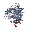
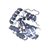
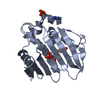
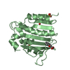
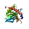
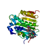
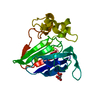
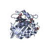
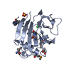
| Entry | Database: PDB / ID: 4urn | ||||||
|---|---|---|---|---|---|---|---|
| Title | Crystal Structure of Staph ParE 24kDa in complex with Novobiocin | ||||||
 Components Components | DNA TOPOISOMERASE IV, B SUBUNIT | ||||||
 Keywords Keywords | ISOMERASE / ANTIBIOTICS / GYRASE / NATURAL PRODUCT | ||||||
| Function / homology | Histidine kinase-like ATPase, C-terminal domain / Heat Shock Protein 90 / 2-Layer Sandwich / Alpha Beta / NOVOBIOCIN / :  Function and homology information Function and homology information | ||||||
| Biological species |   STAPHYLOCOCCUS AUREUS (bacteria) STAPHYLOCOCCUS AUREUS (bacteria) | ||||||
| Method |  X-RAY DIFFRACTION / SYNCHROTRON / MOLECULAR REPLACEMENT / Resolution: 2.3 Å X-RAY DIFFRACTION / SYNCHROTRON / MOLECULAR REPLACEMENT / Resolution: 2.3 Å | ||||||
 Authors Authors | Lu, J. / Patel, S. / Sharma, N. / Soisson, S. / Kishii, R. / Takei, M. / Fukuda, Y. / Lumb, K.J. / Singh, S.B. | ||||||
 Citation Citation | Journal: Acs Chem.Biol. / Year: 2014 Title: Structures of Kibdelomycin Bound to Staphylococcus Aureus Gyrb and Pare Showed a Novel U-Shaped Binding Mode. Authors: Lu, J. / Patel, S. / Sharma, N. / Soisson, S.M. / Kishii, R. / Takei, M. / Fukuda, Y. / Lumb, K.J. / Singh, S.B. | ||||||
| History |
|
- Structure visualization
| Structure viewer | Molecule: MolmilJmol/JSmol |
|---|
- Downloads & links
-Download
| PDBx/mmCIF format | 4urn.cif.gz | 120.4 KB | Display | PDBx/mmCIF format |
|---|---|---|---|---|
| PDB format | pdb4urn.ent.gz | 94 KB | Display | PDB format |
| PDBx/mmJSON format | 4urn.json.gz | Tree view | PDBx/mmJSON format | |
| Others |  Other downloads Other downloads |
-Validation report
| Arichive directory | https://data.pdbj.org/pub/pdb/validation_reports/ur/4urnftp://data.pdbj.org/pub/pdb/validation_reports/ur/4urn | HTTPS FTP |
|---|
-Related structure data
| Related structure data |  4urlC  4urmC  4uroC  1s14S C: citing same article ( S: Starting model for refinement |
|---|---|
| Similar structure data |

-Links
 PDBj
PDBj- Assembly
| Deposited unit | 
| ||||||||
|---|---|---|---|---|---|---|---|---|---|
| 1 | 
| ||||||||
| 2 | 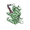
| ||||||||
| 3 | 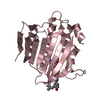
| ||||||||
| Unit cell |
|
-Components
| #1: Protein | Mass: 24796.766 Da / Num. of mol.: 3 / Fragment: N-TERMINAL DOMAIN, RESIDUES 1-225 Source method: isolated from a genetically manipulated source Source: (gene. exp.) STAPHYLOCOCCUS AUREUS (bacteria) / Production host: References: UniProt: X5EN43, Isomerases; Other isomerases; Sole sub-subclass for isomerases that do not belong in the other subclasses #2: Chemical | 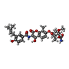  Mass: 612.624 Da / Num. of mol.: 3 / Source method: obtained synthetically / Formula: C31H36N2O11 / Comment: antibiotic*YM Mass: 612.624 Da / Num. of mol.: 3 / Source method: obtained synthetically / Formula: C31H36N2O11 / Comment: antibiotic*YM#3: Water | ChemComp-HOH / |  Mass: 18.015 Da / Num. of mol.: 50 / Source method: isolated from a natural source / Formula: H2O Mass: 18.015 Da / Num. of mol.: 50 / Source method: isolated from a natural source / Formula: H2O |
|---|
-Experimental details
-Experiment
| Experiment | Method: X-RAY DIFFRACTION / Number of used crystals: 1 |
|---|
- Sample preparation
| Crystal | Density Matthews: 2.23 Å3/Da / Density % sol: 44.76 % / Description: NONE |
|---|---|
| Crystal grow | pH: 7.5 Details: 0.15M AMMONIUM ACETATE, 0.075M TRIS PH 8.5 AND 19% PEG 3350 |
-Data collection
| Diffraction | Mean temperature: 293 K |
|---|---|
| Diffraction source | Source: SYNCHROTRON / Site: APS  / Beamline: 17-ID / Wavelength: 1 / Beamline: 17-ID / Wavelength: 1 |
| Detector | Type: DECTRIS PILATUS 6M / Detector: PIXEL / Date: Jun 20, 2008 |
| Radiation | Protocol: SINGLE WAVELENGTH / Monochromatic (M) / Laue (L): M / Scattering type: x-ray |
| Radiation wavelength | Wavelength: 1 Å / Relative weight: 1 |
| Reflection | Resolution: 2.3→50 Å / Num. obs: 28285 / % possible obs: 97 % / Observed criterion σ(I): 3.46 / Redundancy: 4.1 % / Biso Wilson estimate: 48.81 Å2 / Rmerge(I) obs: 0.09 / Net I/σ(I): 10.3 |
| Reflection shell | Resolution: 2.3→2.35 Å / Redundancy: 4.3 % / Rmerge(I) obs: 0.32 / Mean I/σ(I) obs: 3.46 / % possible all: 98.4 |
- Processing
| Software |
| ||||||||||||||||||||||||||||||||||||||||||||||||||||||||||||||||||||||||||||||||||||||||||||||||||||||||||||||||||
|---|---|---|---|---|---|---|---|---|---|---|---|---|---|---|---|---|---|---|---|---|---|---|---|---|---|---|---|---|---|---|---|---|---|---|---|---|---|---|---|---|---|---|---|---|---|---|---|---|---|---|---|---|---|---|---|---|---|---|---|---|---|---|---|---|---|---|---|---|---|---|---|---|---|---|---|---|---|---|---|---|---|---|---|---|---|---|---|---|---|---|---|---|---|---|---|---|---|---|---|---|---|---|---|---|---|---|---|---|---|---|---|---|---|---|---|
| Refinement | Method to determine structure: MOLECULAR REPLACEMENT Starting model: PDB ENTRY 1S14 Resolution: 2.3→19.47 Å / Cor.coef. Fo:Fc: 0.8844 / Cor.coef. Fo:Fc free: 0.8583 / SU R Cruickshank DPI: 0.333 / Cross valid method: THROUGHOUT / σ(F): 0 / SU R Blow DPI: 0.311 / SU Rfree Blow DPI: 0.236 / SU Rfree Cruickshank DPI: 0.245
| ||||||||||||||||||||||||||||||||||||||||||||||||||||||||||||||||||||||||||||||||||||||||||||||||||||||||||||||||||
| Displacement parameters | Biso mean: 50.64 Å2
| ||||||||||||||||||||||||||||||||||||||||||||||||||||||||||||||||||||||||||||||||||||||||||||||||||||||||||||||||||
| Refine analyze | Luzzati coordinate error obs: 0.338 Å | ||||||||||||||||||||||||||||||||||||||||||||||||||||||||||||||||||||||||||||||||||||||||||||||||||||||||||||||||||
| Refinement step | Cycle: LAST / Resolution: 2.3→19.47 Å
| ||||||||||||||||||||||||||||||||||||||||||||||||||||||||||||||||||||||||||||||||||||||||||||||||||||||||||||||||||
| Refine LS restraints |
| ||||||||||||||||||||||||||||||||||||||||||||||||||||||||||||||||||||||||||||||||||||||||||||||||||||||||||||||||||
| LS refinement shell | Resolution: 2.3→2.39 Å / Total num. of bins used: 14
|
