DNA negative supercoiling activity / DNA topoisomerase (ATP-hydrolysing) / DNA topological change / DNA-templated DNA replication / chromosome / response to antibiotic / DNA binding / ATP binding / metal ion binding / cytoplasm Similarity search - Function
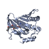
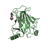
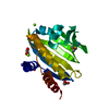
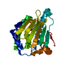
DNA gyrase subunit B, TOPRIM domain / DNA gyrase, subunit B / DNA topoisomerase, type IIA, subunit B / DNA gyrase B subunit, C-terminal / DNA gyrase B subunit, carboxyl terminus / DNA topoisomerase, type IIA, subunit B, domain 2 / DNA gyrase B / DNA topoisomerase, type IIA / DNA topoisomerase, type IIA, conserved site / DNA topoisomerase II signature. ...DNA gyrase subunit B, TOPRIM domain / DNA gyrase, subunit B / DNA topoisomerase, type IIA, subunit B / DNA gyrase B subunit, C-terminal / DNA gyrase B subunit, carboxyl terminus / DNA topoisomerase, type IIA, subunit B, domain 2 / DNA gyrase B / DNA topoisomerase, type IIA / DNA topoisomerase, type IIA, conserved site / DNA topoisomerase II signature. / TopoisomeraseII / DNA topoisomerase, type IIA, subunit B, C-terminal / Toprim domain / DNA topoisomerase, type IIA-like domain superfamily / Toprim domain profile. / TOPRIM domain / Histidine kinase-like ATPase, C-terminal domain / Heat Shock Protein 90 / Histidine kinase-, DNA gyrase B-, and HSP90-like ATPase / Histidine kinase-like ATPases / Histidine kinase/HSP90-like ATPase / Histidine kinase/HSP90-like ATPase superfamily / Ribosomal protein S5 domain 2-type fold, subgroup / Ribosomal protein S5 domain 2-type fold / 2-Layer Sandwich / Alpha Beta Similarity search - Domain/homology
Method to determine structure: OTHER Starting model: NONE Resolution: 2.94→30.44 Å / Cor.coef. Fo:Fc: 0.9051 / Cor.coef. Fo:Fc free: 0.8493 / Cross valid method: THROUGHOUT / σ(F): 0 / SU Rfree Blow DPI: 0.401 Details: IDEAL-DIST CONTACT TERM CONTACT SETUP. ALL ATOMS HAVE CCP4 ATOM TYPE FROM LIBRARY
Rfactor
Num. reflection
% reflection
Selection details
Rfree
0.2563
1012
5.06 %
RANDOM
Rwork
0.1953
-
-
-
obs
0.1984
20004
99.71 %
-
Displacement parameters
Biso mean: 49.38 Å2
Baniso -1
Baniso -2
Baniso -3
1-
5.2479 Å2
0 Å2
-2.3384 Å2
2-
-
8.1216 Å2
0 Å2
3-
-
-
-13.3695 Å2
Refine analyze
Luzzati coordinate error obs: 0.346 Å
Refinement step
Cycle: LAST / Resolution: 2.94→30.44 Å
Protein
Nucleic acid
Ligand
Solvent
Total
Num. atoms
5875
0
256
18
6149
Refine LS restraints
Refine-ID
Type
Dev ideal
Number
Restraint function
Weight
X-RAY DIFFRACTION
t_bond_d
0.01
6253
HARMONIC
2
X-RAY DIFFRACTION
t_angle_deg
1.25
8558
HARMONIC
2
X-RAY DIFFRACTION
t_dihedral_angle_d
2195
SINUSOIDAL
2
X-RAY DIFFRACTION
t_incorr_chiral_ct
X-RAY DIFFRACTION
t_pseud_angle
X-RAY DIFFRACTION
t_trig_c_planes
166
HARMONIC
8
X-RAY DIFFRACTION
t_gen_planes
979
HARMONIC
8
X-RAY DIFFRACTION
t_it
6253
HARMONIC
20
X-RAY DIFFRACTION
t_nbd
X-RAY DIFFRACTION
t_omega_torsion
1.86
X-RAY DIFFRACTION
t_other_torsion
20.53
X-RAY DIFFRACTION
t_improper_torsion
X-RAY DIFFRACTION
t_chiral_improper_torsion
873
SEMIHARMONIC
5
X-RAY DIFFRACTION
t_sum_occupancies
X-RAY DIFFRACTION
t_utility_distance
X-RAY DIFFRACTION
t_utility_angle
X-RAY DIFFRACTION
t_utility_torsion
X-RAY DIFFRACTION
t_ideal_dist_contact
7255
SEMIHARMONIC
4
LS refinement shell
Resolution: 2.94→3.1 Å / Total num. of bins used: 10
Rfactor
Num. reflection
% reflection
Rfree
0.2851
135
4.63 %
Rwork
0.2162
2780
-
all
0.2195
2915
-
obs
-
-
99.71 %
+
About Yorodumi
-
News
-
Feb 9, 2022. New format data for meta-information of EMDB entries
New format data for meta-information of EMDB entries
Version 3 of the EMDB header file is now the official format.
The previous official version 1.9 will be removed from the archive.
In the structure databanks used in Yorodumi, some data are registered as the other names, "COVID-19 virus" and "2019-nCoV". Here are the details of the virus and the list of structure data.
Jan 31, 2019. EMDB accession codes are about to change! (news from PDBe EMDB page)
EMDB accession codes are about to change! (news from PDBe EMDB page)
The allocation of 4 digits for EMDB accession codes will soon come to an end. Whilst these codes will remain in use, new EMDB accession codes will include an additional digit and will expand incrementally as the available range of codes is exhausted. The current 4-digit format prefixed with “EMD-” (i.e. EMD-XXXX) will advance to a 5-digit format (i.e. EMD-XXXXX), and so on. It is currently estimated that the 4-digit codes will be depleted around Spring 2019, at which point the 5-digit format will come into force.
The EM Navigator/Yorodumi systems omit the EMD- prefix.
Related info.:Q: What is EMD? / ID/Accession-code notation in Yorodumi/EM Navigator
Yorodumi is a browser for structure data from EMDB, PDB, SASBDB, etc.
This page is also the successor to EM Navigator detail page, and also detail information page/front-end page for Omokage search.
The word "yorodu" (or yorozu) is an old Japanese word meaning "ten thousand". "mi" (miru) is to see.
Related info.:EMDB / PDB / SASBDB / Comparison of 3 databanks / Yorodumi Search / Aug 31, 2016. New EM Navigator & Yorodumi / Yorodumi Papers / Jmol/JSmol / Function and homology information / Changes in new EM Navigator and Yorodumi
 Movie
Movie Controller

 Yorodumi
 Open data
 Basic information
 Components
 Keywords
 Function and homology information
 STAPHYLOCOCCUS AUREUS (bacteria)
 X-RAY DIFFRACTION /
  Authors
 Citation
 Structure visualization
 Downloads & links
 Other downloads


 PDBj
PDBj


 Assembly
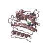
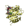
 Mass: 939.872 Da / Num. of mol.: 4 / Source method: obtained synthetically / Formula: C44H60Cl2N4O14
 Mass: 18.015 Da / Num. of mol.: 18 / Source method: isolated from a natural source / Formula: H2O
 Sample preparation
 / Beamline: 22-ID / Wavelength: 1
  Processing