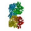
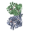
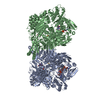
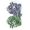
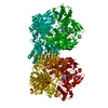
Entry Database : PDB / ID : 4uhxTitle Human aldehyde oxidase in complex with phthalazine and thioridazine ALDEHYDE OXIDASE Keywords / / / / / / Function / homology Function Domain/homology Component
/ / / / / / / / / / / / / / / / / / / / / / / / / / / / / / / / / / / / / / / / / / / / / / / / / / / / / / / / / / / / / / / / / / / / / / / / / / / / / / / / / / / / / / / / / / / / / / / Biological species HOMO SAPIENS (human)Method / / / Resolution : 2.7 Å Authors Coelho, C. / Romao, M.J. / Santos-Silva, T. Journal : Nat.Chem.Biol. / Year : 2015Title : Structural Insights Into Xenobiotic and Inhibitor Binding to Human Aldehyde OxidaseAuthors : Coelho, C. / Foti, A. / Hartmann, T. / Santos-Silva, T. / Leimkuhler, S. / Romao, M.J. History Deposition Mar 26, 2015 Deposition site / Processing site Revision 1.0 Sep 2, 2015 Provider / Type Revision 1.1 Sep 9, 2015 Group Revision 1.2 Sep 30, 2015 Group Revision 1.3 Jan 10, 2024 Group Data collection / Database references ... Data collection / Database references / Derived calculations / Other / Refinement description Category chem_comp_atom / chem_comp_bond ... chem_comp_atom / chem_comp_bond / database_2 / pdbx_database_status / pdbx_initial_refinement_model / struct_site Item _database_2.pdbx_DOI / _database_2.pdbx_database_accession ... _database_2.pdbx_DOI / _database_2.pdbx_database_accession / _pdbx_database_status.status_code_sf / _struct_site.pdbx_auth_asym_id / _struct_site.pdbx_auth_comp_id / _struct_site.pdbx_auth_seq_id
Show all Show less
 Movie
Movie Controller

 Yorodumi
 Open data
 Basic information
 Components
 Keywords
 Function and homology information
 HOMO SAPIENS (human)
 X-RAY DIFFRACTION /
  Authors
 Citation
 Structure visualization
 Downloads & links
 Other downloads


 PDBj
PDBj


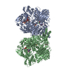
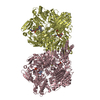
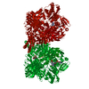
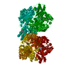
 Assembly
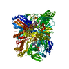


 Mass: 175.820 Da / Num. of mol.: 2 / Source method: obtained synthetically / Formula: Fe2S2
 Mass: 395.352 Da / Num. of mol.: 1 / Source method: obtained synthetically / Formula: C10H14N5O6PS2
 Mass: 161.012 Da / Num. of mol.: 1 / Source method: obtained synthetically / Formula: HMoO2S
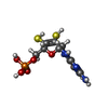
 Mass: 785.550 Da / Num. of mol.: 1 / Source method: obtained synthetically / Formula: C27H33N9O15P2 / Comment: FAD*YM
 Mass: 130.147 Da / Num. of mol.: 1 / Source method: obtained synthetically / Formula: C8H6N2
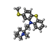
 Mass: 370.575 Da / Num. of mol.: 1 / Source method: obtained synthetically / Formula: C21H26N2S2 / Comment: antipsychotic*YM
 Mass: 370.575 Da / Num. of mol.: 1 / Source method: obtained synthetically / Formula: C21H26N2S2
 Mass: 102.046 Da / Num. of mol.: 2 / Source method: obtained synthetically / Formula: C3H2O4
 Sample preparation
 / Beamline: X06SA / Wavelength: 0.9
  Processing