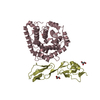
- PDB-4uhr: Thermostabilised HUMAN A2a Receptor with CGS21680 bound -
+
Open data
ID or keywords:
Loading...
-
Basic information
Entry
Database: PDB / ID: 4uhr
Title
Thermostabilised HUMAN A2a Receptor with CGS21680 bound
Components
THERMOSTABILISED HUMAN A2A RECEPTOR
Keywords
SIGNALING PROTEIN / SIGANLING PROTEIN / G PROTEIN COUPLED RECEPTOR / SEVEN-HELIX RECEPTOR / INTEGRAL MEMBRANE PROTEIN / AGONIST BOUND FORM / THERMOSTABILISING POINT MUTATIONS / GPCR / 7TM RECEPTOR
Function / homology
Function and homology information
regulation of norepinephrine secretion / negative regulation of alpha-beta T cell activation / positive regulation of circadian sleep/wake cycle, sleep / Adenosine P1 receptors / positive regulation of acetylcholine secretion, neurotransmission / G protein-coupled adenosine receptor activity / response to purine-containing compound / G protein-coupled adenosine receptor signaling pathway / NGF-independant TRKA activation / Surfactant metabolism ...regulation of norepinephrine secretion / negative regulation of alpha-beta T cell activation / positive regulation of circadian sleep/wake cycle, sleep / Adenosine P1 receptors / positive regulation of acetylcholine secretion, neurotransmission / G protein-coupled adenosine receptor activity / response to purine-containing compound / G protein-coupled adenosine receptor signaling pathway / NGF-independant TRKA activation / Surfactant metabolism / synaptic transmission, dopaminergic / type 5 metabotropic glutamate receptor binding / negative regulation of vascular permeability / synaptic transmission, cholinergic / intermediate filament / presynaptic active zone / positive regulation of urine volume / response to caffeine / blood circulation / sensory perception / positive regulation of glutamate secretion / eating behavior / inhibitory postsynaptic potential / regulation of calcium ion transport / alpha-actinin binding / asymmetric synapse / axolemma / membrane depolarization / cellular defense response / prepulse inhibition / phagocytosis / neuron projection morphogenesis / positive regulation of synaptic transmission, glutamatergic / astrocyte activation / presynaptic modulation of chemical synaptic transmission / positive regulation of long-term synaptic potentiation / positive regulation of synaptic transmission, GABAergic / positive regulation of protein secretion / central nervous system development / response to amphetamine / regulation of mitochondrial membrane potential / positive regulation of apoptotic signaling pathway / apoptotic signaling pathway / synaptic transmission, glutamatergic / excitatory postsynaptic potential / locomotory behavior / negative regulation of inflammatory response / vasodilation / adenylate cyclase-modulating G protein-coupled receptor signaling pathway / blood coagulation / cell-cell signaling / adenylate cyclase-activating G protein-coupled receptor signaling pathway / presynaptic membrane / G alpha (s) signalling events / phospholipase C-activating G protein-coupled receptor signaling pathway / negative regulation of neuron apoptotic process / calmodulin binding / positive regulation of ERK1 and ERK2 cascade / postsynaptic membrane / response to xenobiotic stimulus / inflammatory response / negative regulation of cell population proliferation / neuronal cell body / apoptotic process / regulation of DNA-templated transcription / lipid binding / dendrite / protein-containing complex binding / glutamatergic synapse / enzyme binding / membrane / identical protein binding / plasma membrane Similarity search - Function
Mass: 36022.773 Da / Num. of mol.: 1 / Mutation: YES Source method: isolated from a genetically manipulated source Details: THE CONSTRUCT WAS TRUNCATED AFTER RESIDUE 316 OF THE A2A SEQUENCE, BUT HAS AN 9 RESIDUE SEQUENCE THAT INCLUDES A LINKER AND THE TEV CLEAVAGE SEQUENCE (AAAENLYFQ) AT THE C-TERMINUS Source: (gene. exp.) HOMO SAPIENS (human) / Tissue: BRAIN / Plasmid: PBACPAK8 / Cell line (production host): High Five / Production host: TRICHOPLUSIA NI (cabbage looper) / References: UniProt: P29274
Resolution: 2.6→64.98 Å / Cor.coef. Fo:Fc: 0.909 / Cor.coef. Fo:Fc free: 0.871 / SU B: 13.395 / SU ML: 0.282 / Cross valid method: THROUGHOUT / ESU R: 0.619 / ESU R Free: 0.366 / Stereochemistry target values: MAXIMUM LIKELIHOOD / Details: HYDROGENS HAVE BEEN ADDED IN THE RIDING POSITIONS.
Rfactor
Num. reflection
% reflection
Selection details
Rfree
0.3116
679
5 %
RANDOM
Rwork
0.25868
-
-
-
obs
0.26114
12883
94.75 %
-
Solvent computation
Ion probe radii: 0.8 Å / Shrinkage radii: 0.8 Å / VDW probe radii: 1.2 Å / Solvent model: MASK
 Movie
Movie Controller


 Open data
 Basic information
 Components
 Keywords
 Function and homology information
 HOMO SAPIENS (human)
 X-RAY DIFFRACTION /
  Authors
 Citation
 Structure visualization
 Downloads & links
 Other downloads
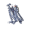

 PDBj
PDBj


 Assembly
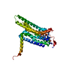
 TRICHOPLUSIA NI (cabbage looper) / References: UniProt: P29274
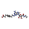
 Mass: 499.520 Da / Num. of mol.: 1 / Source method: obtained synthetically / Formula: C23H29N7O6
 Mass: 18.015 Da / Num. of mol.: 9 / Source method: isolated from a natural source / Formula: H2O
 Sample preparation
 / Beamline: ID23-2 / Wavelength: 0.8726
  Processing