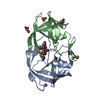
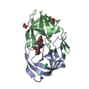
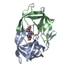
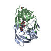
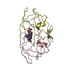
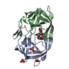
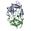
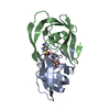
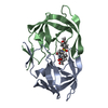
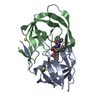
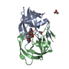
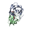
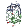
Entry Database : PDB / ID : 4u8wTitle HIV-1 wild Type protease with GRL-050-10A (a Gem-difluoro-bis-Tetrahydrofuran as P2-Ligand) HIV-1 Protease Keywords / / / / / Function / homology Function Domain/homology Component
/ / / / / / / / / / / / / / / / / / / / / / / / / / / / / / / / / / / / / / / / / / / / / / / / / / / / / / / / / / / / / / / / / / / / / / / / / / / / / / / / / / / / / / / / / / / / / / / / / / / Biological species Method / / / Resolution : 1.3 Å Authors Wang, Y.-F. / Agniswamy, J. / Weber, I.T. Funding support Organization Grant number Country National Institutes of Health/National Institute of General Medical Sciences (NIH/NIGMS) GM62920
Journal : Chemmedchem / Year : 2015Title : Design of gem-Difluoro-bis-Tetrahydrofuran as P2 Ligand for HIV-1 Protease Inhibitors to Improve Brain Penetration: Synthesis, X-ray Studies, and Biological Evaluation.Authors : Ghosh, A.K. / Yashchuk, S. / Mizuno, A. / Chakraborty, N. / Agniswamy, J. / Wang, Y.F. / Aoki, M. / Gomez, P.M. / Amano, M. / Weber, I.T. / Mitsuya, H. History Deposition Aug 5, 2014 Deposition site / Processing site Revision 1.0 Nov 5, 2014 Provider / Type Revision 1.1 Dec 31, 2014 Group Revision 1.2 Sep 27, 2017 Group Author supporting evidence / Derived calculations ... Author supporting evidence / Derived calculations / Other / Refinement description / Source and taxonomy Category entity_src_gen / pdbx_audit_support ... entity_src_gen / pdbx_audit_support / pdbx_database_status / pdbx_struct_oper_list / refine / software Item _entity_src_gen.pdbx_alt_source_flag / _pdbx_audit_support.funding_organization ... _entity_src_gen.pdbx_alt_source_flag / _pdbx_audit_support.funding_organization / _pdbx_database_status.pdb_format_compatible / _pdbx_struct_oper_list.symmetry_operation / _refine.pdbx_method_to_determine_struct Revision 1.3 Dec 25, 2019 Group / Category / Item Revision 1.4 Sep 27, 2023 Group Data collection / Database references ... Data collection / Database references / Derived calculations / Refinement description Category chem_comp_atom / chem_comp_bond ... chem_comp_atom / chem_comp_bond / database_2 / pdbx_initial_refinement_model / pdbx_struct_conn_angle / refine_hist / struct_conn Item _database_2.pdbx_DOI / _database_2.pdbx_database_accession ... _database_2.pdbx_DOI / _database_2.pdbx_database_accession / _pdbx_struct_conn_angle.ptnr1_auth_seq_id / _pdbx_struct_conn_angle.ptnr3_auth_seq_id / _pdbx_struct_conn_angle.value / _refine_hist.number_atoms_total / _struct_conn.pdbx_dist_value / _struct_conn.ptnr2_auth_seq_id
Show all Show less
 Movie
Movie Controller

 Yorodumi
 Open data
 Basic information
 Components
 Keywords
 Function and homology information
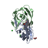
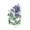
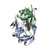
 Human immunodeficiency virus type 1 BH10
 X-RAY DIFFRACTION /
  Authors
 United States, 1items
  Citation
 Structure visualization
 Downloads & links
 Other downloads
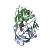


 PDBj
PDBj


 Assembly
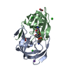

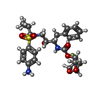


 Mass: 583.645 Da / Num. of mol.: 1 / Source method: obtained synthetically / Formula: C27H35F2N3O7S
 Mass: 22.990 Da / Num. of mol.: 1 / Source method: obtained synthetically / Formula: Na
 Mass: 35.453 Da / Num. of mol.: 3 / Source method: obtained synthetically / Formula: Cl
 Mass: 59.044 Da / Num. of mol.: 2 / Source method: obtained synthetically / Formula: C2H3O2
 Sample preparation
 Processing