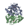
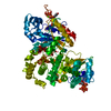
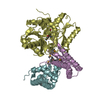
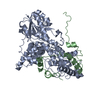
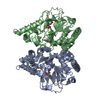
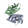
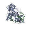
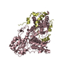
Entry Database : PDB / ID : 4u8hTitle Crystal Structure of Mammalian Period-Cryptochrome Complex Cryptochrome-2 Period circadian protein homolog 2 Keywords / / / Function / homology Function Domain/homology Component
/ / / / / / / / / / / / / / / / / / / / / / / / / / / / / / / / / / / / / / / / / / / / / / / / / / / / / / / / / / / / / / / / / / / / / / / / / / / / / / / / / / / / / / / / / / / / / / / / / / / / / / / / / / / / / / / / / Biological species Mus musculus (house mouse)Method / / / Resolution : 2.798 Å Authors Nangle, S.N. / Rosensweig, C. / Koike, N. / Tei, H. / Takahashi, J.S. / Green, C.B. / Zheng, N. Funding support Organization Grant number Country National Institutes of Health/National Cancer Institute (NIH/NCI) R01-CA107134 Howard Hughes Medical Institute (HHMI) National Institutes of Health/National Institute of General Medical Sciences (NIH/NIGMS) T32-GM007750
Journal : Elife / Year : 2014Title : Molecular assembly of the period-cryptochrome circadian transcriptional repressor complex.Authors : Nangle, S.N. / Rosensweig, C. / Koike, N. / Tei, H. / Takahashi, J.S. / Green, C.B. / Zheng, N. History Deposition Aug 3, 2014 Deposition site / Processing site Revision 1.0 Oct 1, 2014 Provider / Type Revision 1.1 Sep 20, 2017 Group Author supporting evidence / Derived calculations ... Author supporting evidence / Derived calculations / Other / Source and taxonomy Category entity_src_gen / pdbx_audit_support ... entity_src_gen / pdbx_audit_support / pdbx_database_status / pdbx_struct_oper_list Item _entity_src_gen.pdbx_alt_source_flag / _pdbx_audit_support.funding_organization ... _entity_src_gen.pdbx_alt_source_flag / _pdbx_audit_support.funding_organization / _pdbx_database_status.pdb_format_compatible / _pdbx_struct_oper_list.symmetry_operation Revision 1.2 Nov 20, 2019 Group / Category / Item Revision 1.3 Sep 27, 2023 Group Data collection / Database references ... Data collection / Database references / Derived calculations / Refinement description Category chem_comp_atom / chem_comp_bond ... chem_comp_atom / chem_comp_bond / database_2 / pdbx_initial_refinement_model / pdbx_struct_conn_angle / refine_hist / struct_conn / struct_conn_type Item _database_2.pdbx_DOI / _database_2.pdbx_database_accession ... _database_2.pdbx_DOI / _database_2.pdbx_database_accession / _pdbx_struct_conn_angle.ptnr1_auth_asym_id / _pdbx_struct_conn_angle.ptnr1_auth_comp_id / _pdbx_struct_conn_angle.ptnr1_auth_seq_id / _pdbx_struct_conn_angle.ptnr1_label_asym_id / _pdbx_struct_conn_angle.ptnr1_label_atom_id / _pdbx_struct_conn_angle.ptnr1_label_comp_id / _pdbx_struct_conn_angle.ptnr1_label_seq_id / _pdbx_struct_conn_angle.ptnr2_symmetry / _pdbx_struct_conn_angle.ptnr3_auth_asym_id / _pdbx_struct_conn_angle.ptnr3_auth_comp_id / _pdbx_struct_conn_angle.ptnr3_auth_seq_id / _pdbx_struct_conn_angle.ptnr3_label_asym_id / _pdbx_struct_conn_angle.ptnr3_label_atom_id / _pdbx_struct_conn_angle.ptnr3_label_comp_id / _pdbx_struct_conn_angle.ptnr3_label_seq_id / _pdbx_struct_conn_angle.value / _struct_conn.conn_type_id / _struct_conn.id / _struct_conn.pdbx_dist_value / _struct_conn.pdbx_leaving_atom_flag / _struct_conn.ptnr1_auth_asym_id / _struct_conn.ptnr1_auth_comp_id / _struct_conn.ptnr1_auth_seq_id / _struct_conn.ptnr1_label_asym_id / _struct_conn.ptnr1_label_atom_id / _struct_conn.ptnr1_label_comp_id / _struct_conn.ptnr1_label_seq_id / _struct_conn.ptnr2_auth_asym_id / _struct_conn.ptnr2_auth_comp_id / _struct_conn.ptnr2_auth_seq_id / _struct_conn.ptnr2_label_asym_id / _struct_conn.ptnr2_label_atom_id / _struct_conn.ptnr2_label_comp_id / _struct_conn.ptnr2_label_seq_id / _struct_conn.ptnr2_symmetry / _struct_conn_type.id Revision 1.4 Oct 23, 2024 Group / Category / pdbx_modification_feature
Show all Show less
 Movie
Movie Controller


 Open data
 Basic information
 Components
 Keywords
 Function and homology information
 X-RAY DIFFRACTION /
  Authors
 United States, 3items
  Citation
 Structure visualization
 Downloads & links
 Other downloads
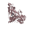


 PDBj
PDBj


 Assembly

 Spodoptera frugiperda (fall armyworm) / References: UniProt: Q9R194
 Mass: 65.409 Da / Num. of mol.: 2 / Source method: obtained synthetically / Formula: Zn
 Mass: 18.015 Da / Num. of mol.: 48 / Source method: isolated from a natural source / Formula: H2O
 Sample preparation
 Processing