 Movie
Movie Controller

[English] 日本語
 Yorodumi
- PDB-4u66: Induced Dimer Structure of Methionine Sulfoxide Reductase U16C fr... -
+ Open data

- Basic information
| Entry | Database: PDB / ID: 4u66 | ||||||
|---|---|---|---|---|---|---|---|
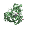
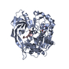
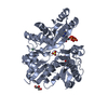
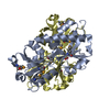
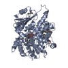
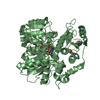
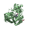
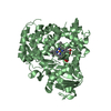
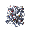
| Title | Induced Dimer Structure of Methionine Sulfoxide Reductase U16C from Clostridium Oremlandii | ||||||
 Components Components | Peptide methionine sulfoxide reductase MsrA | ||||||
 Keywords Keywords | OXIDOREDUCTASE / ALPHA/BETA FOLD / PEPTIDE-METHIONINE (S)-S-OXIDE REDUCTASE | ||||||
| Function / homology |  Function and homology information Function and homology informationL-methionine (S)-S-oxide reductase activity / peptide-methionine (S)-S-oxide reductase / peptide-methionine (S)-S-oxide reductase activity Similarity search - Function | ||||||
| Biological species |  Alkaliphilus oremlandii OhILAs (bacteria) Alkaliphilus oremlandii OhILAs (bacteria) | ||||||
| Method |  X-RAY DIFFRACTION / SYNCHROTRON / MOLECULAR REPLACEMENT / Resolution: 2.9 Å X-RAY DIFFRACTION / SYNCHROTRON / MOLECULAR REPLACEMENT / Resolution: 2.9 Å | ||||||
 Authors Authors | Hwang, K.Y. / Lee, E.H. | ||||||
 Citation Citation | Journal: Plos One / Year: 2015 Title: Evidence for the Dimerization-Mediated Catalysis of Methionine Sulfoxide Reductase A from Clostridium oremlandii Authors: Lee, E.H. / Lee, K. / Kwak, G.H. / Park, Y.S. / Lee, K.J. / Hwang, K.Y. / Kim, H.Y. | ||||||
| History |
|
- Structure visualization
| Structure viewer | Molecule: MolmilJmol/JSmol |
|---|
- Downloads & links
-Download
| PDBx/mmCIF format | 4u66.cif.gz | 133.4 KB | Display | PDBx/mmCIF format |
|---|---|---|---|---|
| PDB format | pdb4u66.ent.gz | 104.2 KB | Display | PDB format |
| PDBx/mmJSON format | 4u66.json.gz | Tree view | PDBx/mmJSON format | |
| Others |  Other downloads Other downloads |
-Validation report
| Arichive directory | https://data.pdbj.org/pub/pdb/validation_reports/u6/4u66ftp://data.pdbj.org/pub/pdb/validation_reports/u6/4u66 | HTTPS FTP |
|---|
-Related structure data
| Related structure data | 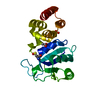 4lwjS S: Starting model for refinement |
|---|---|
| Similar structure data |

-Links
 PDBj
PDBj- Assembly
| Deposited unit | 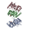
| ||||||||
|---|---|---|---|---|---|---|---|---|---|
| 1 | 
| ||||||||
| 2 | 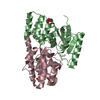
| ||||||||
| Unit cell |
|
-Components
| #1: Protein | Mass: 23729.600 Da / Num. of mol.: 3 / Mutation: U16C Source method: isolated from a genetically manipulated source Source: (gene. exp.) Alkaliphilus oremlandii OhILAs (bacteria)Gene: msrA, Clos_1947 / Plasmid: PET21B / Production host: References: UniProt: A8MI53, peptide-methionine (S)-S-oxide reductase #2: Chemical |   Mass: 96.063 Da / Num. of mol.: 3 / Source method: obtained synthetically / Formula: SO4 Mass: 96.063 Da / Num. of mol.: 3 / Source method: obtained synthetically / Formula: SO4#3: Water | ChemComp-HOH / |  Mass: 18.015 Da / Num. of mol.: 48 / Source method: isolated from a natural source / Formula: H2O Mass: 18.015 Da / Num. of mol.: 48 / Source method: isolated from a natural source / Formula: H2O |
|---|
-Experimental details
-Experiment
| Experiment | Method: X-RAY DIFFRACTION / Number of used crystals: 1 |
|---|
- Sample preparation
| Crystal | Density Matthews: 4.21 Å3/Da / Density % sol: 70.76 % |
|---|---|
| Crystal grow | Temperature: 293 K / Method: vapor diffusion, sitting drop / pH: 7.3 Details: 0.1M TRIS-HCL, 1.2M AMMONIUM SULFATE, 12%(V/V) GLYCEROL |
-Data collection
| Diffraction | Mean temperature: 100 K |
|---|---|
| Diffraction source | Source: SYNCHROTRON / Site: Photon Factory  / Beamline: BL-1A / Wavelength: 1 Å / Beamline: BL-1A / Wavelength: 1 Å |
| Detector | Type: ADSC QUANTUM 4 / Detector: CCD / Date: May 20, 2011 |
| Radiation | Monochromator: SI / Protocol: SINGLE WAVELENGTH / Monochromatic (M) / Laue (L): M / Scattering type: x-ray |
| Radiation wavelength | Wavelength: 1 Å / Relative weight: 1 |
| Reflection | Resolution: 2.9→30 Å / Num. obs: 26615 / % possible obs: 94.6 % / Observed criterion σ(I): 2 / Redundancy: 2.9 % / Biso Wilson estimate: 50.47 Å2 / Net I/σ(I): 11.6 |
| Reflection shell | Resolution: 2.9→2.95 Å / Rejects: 0 / % possible all: 88.6 |
- Processing
| Software |
| ||||||||||||||||||||||||||||||||||||||||||||||||||||||||||||||||||||||||||||||||||||||||||||||||||
|---|---|---|---|---|---|---|---|---|---|---|---|---|---|---|---|---|---|---|---|---|---|---|---|---|---|---|---|---|---|---|---|---|---|---|---|---|---|---|---|---|---|---|---|---|---|---|---|---|---|---|---|---|---|---|---|---|---|---|---|---|---|---|---|---|---|---|---|---|---|---|---|---|---|---|---|---|---|---|---|---|---|---|---|---|---|---|---|---|---|---|---|---|---|---|---|---|---|---|---|
| Refinement | Method to determine structure: MOLECULAR REPLACEMENT Starting model: PDB DNTRY 4LWJ Resolution: 2.9→29.82 Å / SU ML: 0.59 / Cross valid method: FREE R-VALUE / σ(F): 0.2 / Phase error: 28.74 / Stereochemistry target values: ML
| ||||||||||||||||||||||||||||||||||||||||||||||||||||||||||||||||||||||||||||||||||||||||||||||||||
| Solvent computation | Shrinkage radii: 0.9 Å / VDW probe radii: 1.11 Å / Solvent model: FLAT BULK SOLVENT MODEL | ||||||||||||||||||||||||||||||||||||||||||||||||||||||||||||||||||||||||||||||||||||||||||||||||||
| Displacement parameters | Biso max: 172.73 Å2 / Biso mean: 77.93 Å2 / Biso min: 24.39 Å2 | ||||||||||||||||||||||||||||||||||||||||||||||||||||||||||||||||||||||||||||||||||||||||||||||||||
| Refinement step | Cycle: final / Resolution: 2.9→29.82 Å
| ||||||||||||||||||||||||||||||||||||||||||||||||||||||||||||||||||||||||||||||||||||||||||||||||||
| LS refinement shell | Refine-ID: X-RAY DIFFRACTION / Total num. of bins used: 13
|
