Component-ID: _ / Beg auth comp-ID: SER / Beg label comp-ID: SER / End auth comp-ID: ALA / End label comp-ID: ALA / Refine code: _ / Auth seq-ID: 25 - 762 / Label seq-ID: 6 - 743
Dom-ID
Ens-ID
Auth asym-ID
Label asym-ID
1
1
A
A
2
1
B
B
1
2
A
A
2
2
C
C
1
3
A
A
2
3
D
D
1
4
B
B
2
4
C
C
1
5
B
B
2
5
D
D
1
6
C
C
2
6
D
D
NCS ensembles :
ID
1
2
3
4
5
6
-
Components
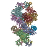
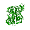
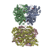
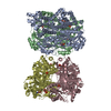
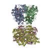
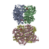
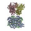
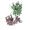
#1: Protein
ATP-dependent6-phosphofructokinase, platelettype / PFK-P / 6-phosphofructokinase type C / Phosphofructo-1-kinase isozyme C / PFK-C / Phosphohexokinase
Mass: 81111.844 Da / Num. of mol.: 4 / Fragment: UNP residues 26-762 Source method: isolated from a genetically manipulated source Source: (gene. exp.) Homo sapiens (human) / Gene: PFKP, PFKF / Plasmid: pET51b / Production host: Escherichia coli BL21(DE3) (bacteria) / Variant (production host): Rosetta pLysS / References: UniProt: Q01813, 6-phosphofructokinase
 Movie
Movie Controller

 Yorodumi
 Open data
 Basic information
 Components
 Keywords
 Function and homology information
 Homo sapiens (human)
 X-RAY DIFFRACTION /
  Authors
 Germany, 1items
  Citation
 Structure visualization
 Downloads & links
 Other downloads


 PDBj
PDBj

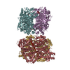
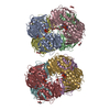
 Assembly
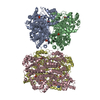


 Mass: 507.181 Da / Num. of mol.: 4 / Source method: obtained synthetically / Formula: C10H16N5O13P3 / Comment: ATP, energy-carrying molecule*YM
 Mass: 94.971 Da / Num. of mol.: 12 / Source method: obtained synthetically / Formula: PO4
 Sample preparation
 Processing