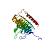
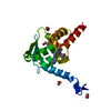
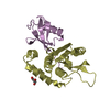
TypeIIIiodothyroninedeiodinase / 5DIII / DIOIII / Type 3 DI / Type-III 5'-deiodinase
Mass: 21696.639 Da / Num. of mol.: 1 / Mutation: yes Source method: isolated from a genetically manipulated source Details: six N-terminal residues are cloning artifact; three C-terminal residues not visible in electron density Source: (gene. exp.) Mus musculus (house mouse) / Gene: Dio3 / Plasmid: pET151 TOPO / Production host: Escherichia coli (E. coli) / References: UniProt: Q91ZI8, EC: 1.97.1.11
Protocol: SINGLE WAVELENGTH / Monochromatic (M) / Laue (L): M / Scattering type: x-ray
Radiation wavelength
Wavelength: 0.9795 Å / Relative weight: 1
Reflection
Resolution: 1.9→20 Å / Num. all: 14304 / Num. obs: 14015 / % possible obs: 96 % / Redundancy: 3.7 % / Rmerge(I) obs: 0.075 / Net I/σ(I): 13.9
-
Processing
Software
Name
Version
Classification
REFMAC
5.5.0042
refinement
PDB_EXTRACT
3.14
dataextraction
XDS
datareduction
SHELX
phasing
ARP
modelbuilding
Coot
modelbuilding
Refinement
Method to determine structure: SAD / Resolution: 1.9→42.07 Å / Cor.coef. Fo:Fc: 0.93 / Cor.coef. Fo:Fc free: 0.9 / SU B: 3.035 / SU ML: 0.09 / Cross valid method: THROUGHOUT / σ(F): 0 / ESU R: 0.171 / ESU R Free: 0.152 / Stereochemistry target values: MAXIMUM LIKELIHOOD Details: HYDROGENS HAVE BEEN ADDED IN THE RIDING POSITIONS U VALUES : REFINED INDIVIDUALLY
Rfactor
Num. reflection
% reflection
Selection details
Rfree
0.2272
706
5 %
RANDOM
Rwork
0.1866
13309
-
-
obs
0.1885
14015
95.95 %
-
Solvent computation
Ion probe radii: 0.8 Å / Shrinkage radii: 0.8 Å / VDW probe radii: 1.2 Å / Solvent model: BABINET MODEL WITH MASK
In the structure databanks used in Yorodumi, some data are registered as the other names, "COVID-19 virus" and "2019-nCoV". Here are the details of the virus and the list of structure data.
Jan 31, 2019. EMDB accession codes are about to change! (news from PDBe EMDB page)
EMDB accession codes are about to change! (news from PDBe EMDB page)
The allocation of 4 digits for EMDB accession codes will soon come to an end. Whilst these codes will remain in use, new EMDB accession codes will include an additional digit and will expand incrementally as the available range of codes is exhausted. The current 4-digit format prefixed with “EMD-” (i.e. EMD-XXXX) will advance to a 5-digit format (i.e. EMD-XXXXX), and so on. It is currently estimated that the 4-digit codes will be depleted around Spring 2019, at which point the 5-digit format will come into force.
The EM Navigator/Yorodumi systems omit the EMD- prefix.
Related info.:Q: What is EMD? / ID/Accession-code notation in Yorodumi/EM Navigator
Yorodumi is a browser for structure data from EMDB, PDB, SASBDB, etc.
This page is also the successor to EM Navigator detail page, and also detail information page/front-end page for Omokage search.
The word "yorodu" (or yorozu) is an old Japanese word meaning "ten thousand". "mi" (miru) is to see.
Related info.:EMDB / PDB / SASBDB / Comparison of 3 databanks / Yorodumi Search / Aug 31, 2016. New EM Navigator & Yorodumi / Yorodumi Papers / Jmol/JSmol / Function and homology information / Changes in new EM Navigator and Yorodumi
 Movie
Movie Controller

 Yorodumi
 Open data
 Basic information
 Components
 Keywords
 Function and homology information
 X-RAY DIFFRACTION /
  Authors
 Citation
 Structure visualization
 Downloads & links
 Other downloads
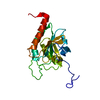


 PDBj
PDBj Assembly
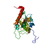

 Mass: 18.015 Da / Num. of mol.: 93 / Source method: isolated from a natural source / Formula: H2O
 Sample preparation
 / Beamline: ID14-4 / Wavelength: 0.9795 Å
 Processing