 Movie
Movie Controller


+ Open data

- Basic information
| Entry | Database: PDB / ID: 4s2k | ||||||
|---|---|---|---|---|---|---|---|
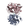
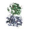
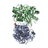
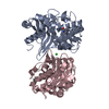
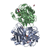
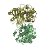
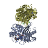
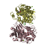
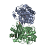
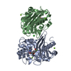
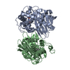
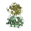
| Title | OXA-48 in complex with Avibactam at pH 7.5 | ||||||
 Components Components | Beta-lactamase | ||||||
 Keywords Keywords | hydrolase/antibiotic / inhibitor / beta-lactamase / hydrolase-antibiotic complex | ||||||
| Function / homology |  Function and homology information Function and homology informationpenicillin binding / antibiotic catabolic process / cell wall organization / beta-lactamase activity / beta-lactamase / response to antibiotic / plasma membrane Similarity search - Function | ||||||
| Biological species |  Klebsiella pneumoniae (bacteria) Klebsiella pneumoniae (bacteria) | ||||||
| Method |  X-RAY DIFFRACTION / SYNCHROTRON / MOLECULAR REPLACEMENT / Resolution: 2.1 Å X-RAY DIFFRACTION / SYNCHROTRON / MOLECULAR REPLACEMENT / Resolution: 2.1 Å | ||||||
 Authors Authors | King, D.T. / Strynadka, N.C.J. | ||||||
 Citation Citation | Journal: ACS Infect Dis / Year: 2015 Title: Molecular Mechanism of Avibactam-Mediated beta-Lactamase Inhibition. Authors: King, D.T. / King, A.M. / Lal, S.M. / Wright, G.D. / Strynadka, N.C. | ||||||
| History |
|
- Structure visualization
| Structure viewer | Molecule: MolmilJmol/JSmol |
|---|
- Downloads & links
-Download
| PDBx/mmCIF format | 4s2k.cif.gz | 407.8 KB | Display | PDBx/mmCIF format |
|---|---|---|---|---|
| PDB format | pdb4s2k.ent.gz | 337.4 KB | Display | PDB format |
| PDBx/mmJSON format | 4s2k.json.gz | Tree view | PDBx/mmJSON format | |
| Others |  Other downloads Other downloads |
-Validation report
| Summary document | 4s2k_validation.pdf.gz | 1.6 MB | Display | wwPDB validaton report |
|---|---|---|---|---|
| Full document | 4s2k_full_validation.pdf.gz | 1.6 MB | Display | |
| Data in XML | 4s2k_validation.xml.gz | 40.5 KB | Display | |
| Data in CIF | 4s2k_validation.cif.gz | 57.8 KB | Display | |
| Arichive directory | https://data.pdbj.org/pub/pdb/validation_reports/s2/4s2kftp://data.pdbj.org/pub/pdb/validation_reports/s2/4s2k | HTTPS FTP |
-Related structure data
| Related structure data | 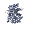 4s2iC  4s2jC 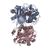 4s2nC 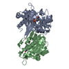 4s2oC 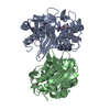 4s2pC  3hbrS S: Starting model for refinement C: citing same article ( |
|---|---|
| Similar structure data |


-Links
 PDBj
PDBj- Assembly
| Deposited unit | 
| ||||||||
|---|---|---|---|---|---|---|---|---|---|
| 1 | 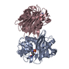
| ||||||||
| 2 | 
| ||||||||
| 3 | 
| ||||||||
| 4 | 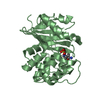
| ||||||||
| 5 | 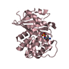
| ||||||||
| 6 | 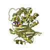
| ||||||||
| Unit cell |
| ||||||||
| Details | Authors report that the protein behaves as a dimer in solution as determined by gel filtration |
-Components
| #1: Protein | Mass: 30396.721 Da / Num. of mol.: 4 Source method: isolated from a genetically manipulated source Source: (gene. exp.) Klebsiella pneumoniae (bacteria) / Gene: bla OXA-48, blaOXA-48, FP68_27275, KPE71T_00045 / Production host: #2: Chemical | ChemComp-NXL / (   Mass: 267.260 Da / Num. of mol.: 4 / Source method: obtained synthetically / Formula: C7H13N3O6S / Comment: antibiotic*YM Mass: 267.260 Da / Num. of mol.: 4 / Source method: obtained synthetically / Formula: C7H13N3O6S / Comment: antibiotic*YM#3: Water | ChemComp-HOH / |  Mass: 18.015 Da / Num. of mol.: 537 / Source method: isolated from a natural source / Formula: H2O Mass: 18.015 Da / Num. of mol.: 537 / Source method: isolated from a natural source / Formula: H2OHas protein modification | Y | |
|---|
-Experimental details
-Experiment
| Experiment | Method: X-RAY DIFFRACTION / Number of used crystals: 1 |
|---|
- Sample preparation
| Crystal | Density Matthews: 2.54 Å3/Da / Density % sol: 51.52 % |
|---|---|
| Crystal grow | Temperature: 298.15 K / Method: vapor diffusion, sitting drop / pH: 7.5 Details: 200mM tris pH 7.5, 0.1M ammonium chloride, 40% MPD, 5% PEG 8K, 100 mM NaCl, 2 mM avibactam, VAPOR DIFFUSION, SITTING DROP, temperature 298.15K |
-Data collection
| Diffraction | Mean temperature: 100 K |
|---|---|
| Diffraction source | Source: SYNCHROTRON / Site: CLSI  / Beamline: 08B1-1 / Wavelength: 1 Å / Beamline: 08B1-1 / Wavelength: 1 Å |
| Detector | Type: RAYONIX MX300HE / Detector: CCD / Date: Jun 13, 2013 |
| Radiation | Monochromator: double crystal monochromator (DCM) / Protocol: SINGLE WAVELENGTH / Monochromatic (M) / Laue (L): M / Scattering type: x-ray |
| Radiation wavelength | Wavelength: 1 Å / Relative weight: 1 |
| Reflection | Resolution: 2.1→52.42 Å / Num. all: 69789 / Num. obs: 69650 / % possible obs: 99.8 % / Biso Wilson estimate: 28.8 Å2 |
| Reflection shell | Resolution: 2.1→2.21 Å / % possible all: 100 |
- Processing
| Software |
| |||||||||||||||||||||||||||||||||||||||||||||||||||||||||||||||||||||||||||||||||||||||||||||||||||||||||||||||||||||||||||||
|---|---|---|---|---|---|---|---|---|---|---|---|---|---|---|---|---|---|---|---|---|---|---|---|---|---|---|---|---|---|---|---|---|---|---|---|---|---|---|---|---|---|---|---|---|---|---|---|---|---|---|---|---|---|---|---|---|---|---|---|---|---|---|---|---|---|---|---|---|---|---|---|---|---|---|---|---|---|---|---|---|---|---|---|---|---|---|---|---|---|---|---|---|---|---|---|---|---|---|---|---|---|---|---|---|---|---|---|---|---|---|---|---|---|---|---|---|---|---|---|---|---|---|---|---|---|---|
| Refinement | Method to determine structure: MOLECULAR REPLACEMENT Starting model: 3HBR Resolution: 2.1→42.25 Å / Cor.coef. Fo:Fc: 0.9384 / Cor.coef. Fo:Fc free: 0.9198 / SU R Cruickshank DPI: 0.18 / Cross valid method: THROUGHOUT / σ(F): 0
| |||||||||||||||||||||||||||||||||||||||||||||||||||||||||||||||||||||||||||||||||||||||||||||||||||||||||||||||||||||||||||||
| Displacement parameters | Biso mean: 30.42 Å2
| |||||||||||||||||||||||||||||||||||||||||||||||||||||||||||||||||||||||||||||||||||||||||||||||||||||||||||||||||||||||||||||
| Refine analyze | Luzzati coordinate error obs: 0.253 Å | |||||||||||||||||||||||||||||||||||||||||||||||||||||||||||||||||||||||||||||||||||||||||||||||||||||||||||||||||||||||||||||
| Refinement step | Cycle: LAST / Resolution: 2.1→42.25 Å
| |||||||||||||||||||||||||||||||||||||||||||||||||||||||||||||||||||||||||||||||||||||||||||||||||||||||||||||||||||||||||||||
| Refine LS restraints |
| |||||||||||||||||||||||||||||||||||||||||||||||||||||||||||||||||||||||||||||||||||||||||||||||||||||||||||||||||||||||||||||
| LS refinement shell | Resolution: 2.1→2.15 Å / Total num. of bins used: 20
| |||||||||||||||||||||||||||||||||||||||||||||||||||||||||||||||||||||||||||||||||||||||||||||||||||||||||||||||||||||||||||||
| Refinement TLS params. | Method: refined / Refine-ID: X-RAY DIFFRACTION
| |||||||||||||||||||||||||||||||||||||||||||||||||||||||||||||||||||||||||||||||||||||||||||||||||||||||||||||||||||||||||||||
| Refinement TLS group |
|
