 Movie
Movie Controller

[English] 日本語
 Yorodumi
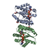
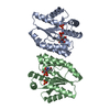
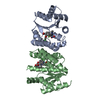
- PDB-4s2e: Phosphate ion bound Crystal structure of thymidylate kinase (aq_9... -
+ Open data

- Basic information
| Entry | Database: PDB / ID: 4s2e | ||||||
|---|---|---|---|---|---|---|---|
| Title | Phosphate ion bound Crystal structure of thymidylate kinase (aq_969) from Aquifex Aeolicus VF5 | ||||||
 Components Components | Thymidylate kinase | ||||||
 Keywords Keywords | TRANSFERASE / ATP binding / TMP binding | ||||||
| Function / homology |  Function and homology information Function and homology informationdTMP kinase / dUDP biosynthetic process / dTDP biosynthetic process / dTMP kinase activity / dTTP biosynthetic process / ATP binding / cytoplasm / cytosol Similarity search - Function | ||||||
| Biological species |   Aquifex aeolicus (bacteria) Aquifex aeolicus (bacteria) | ||||||
| Method |  X-RAY DIFFRACTION / MOLECULAR REPLACEMENT / Resolution: 2.35 Å X-RAY DIFFRACTION / MOLECULAR REPLACEMENT / Resolution: 2.35 Å | ||||||
 Authors Authors | Biswas, A. / Jeyakanthan, J. / Sekar, K. | ||||||
 Citation Citation | Journal: Febs J. / Year: 2017 Title: Structural studies of a hyperthermophilic thymidylate kinase enzyme reveal conformational substates along the reaction coordinate. Authors: Biswas, A. / Shukla, A. / Chaudhary, S.K. / Santhosh, R. / Jeyakanthan, J. / Sekar, K. | ||||||
| History |
|
- Structure visualization
| Structure viewer | Molecule: MolmilJmol/JSmol |
|---|
- Downloads & links
-Download
| PDBx/mmCIF format | 4s2e.cif.gz | 87.1 KB | Display | PDBx/mmCIF format |
|---|---|---|---|---|
| PDB format | pdb4s2e.ent.gz | 66.6 KB | Display | PDB format |
| PDBx/mmJSON format | 4s2e.json.gz | Tree view | PDBx/mmJSON format | |
| Others |  Other downloads Other downloads |
-Validation report
| Arichive directory | https://data.pdbj.org/pub/pdb/validation_reports/s2/4s2eftp://data.pdbj.org/pub/pdb/validation_reports/s2/4s2e | HTTPS FTP |
|---|
-Related structure data
| Related structure data | 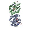 4s35C 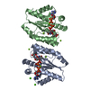 5h56C 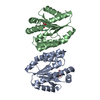 5h5bC  5h5kC  5xaiC 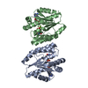 5xb2C  5xb3C  5xb5C 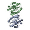 5xbhC 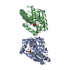 2pbrS S: Starting model for refinement C: citing same article ( |
|---|---|
| Similar structure data |


-Links
 PDBj
PDBj- Assembly
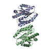
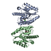
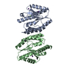
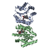
| Deposited unit | 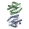
| ||||||||||||
|---|---|---|---|---|---|---|---|---|---|---|---|---|---|
| 1 |
| ||||||||||||
| Unit cell |
| ||||||||||||
| Noncrystallographic symmetry (NCS) | NCS oper:
|
-Components
| #1: Protein | Mass: 22402.961 Da / Num. of mol.: 2 Source method: isolated from a genetically manipulated source Source: (gene. exp.) Aquifex aeolicus (bacteria) / Strain: VF5 / Gene: aq_969, tmk / Plasmid: pET21a / Production host: #2: Chemical |   Mass: 94.971 Da / Num. of mol.: 2 / Source method: obtained synthetically / Formula: PO4 Mass: 94.971 Da / Num. of mol.: 2 / Source method: obtained synthetically / Formula: PO4#3: Water | ChemComp-HOH / |  Mass: 18.015 Da / Num. of mol.: 54 / Source method: isolated from a natural source / Formula: H2O Mass: 18.015 Da / Num. of mol.: 54 / Source method: isolated from a natural source / Formula: H2O |
|---|
-Experimental details
-Experiment
| Experiment | Method: X-RAY DIFFRACTION / Number of used crystals: 1 |
|---|
- Sample preparation
| Crystal | Density Matthews: 2.44 Å3/Da / Density % sol: 49.65 % |
|---|---|
| Crystal grow | Temperature: 293 K / Method: evaporation / pH: 7.5 Details: 0.1 M HEPES, 10% w/v Polyethylene glycol 8,000, 8% v/v Ethylene glycol , Microbatch Underoil, pH 7.5, EVAPORATION, temperature 293K |
-Data collection
| Diffraction | Mean temperature: 100 K |
|---|---|
| Diffraction source | Source: ROTATING ANODE / Type: BRUKER AXS MICROSTAR / Wavelength: 1.5418 Å |
| Detector | Type: MAR scanner 345 mm plate / Detector: IMAGE PLATE / Details: Mirrors |
| Radiation | Monochromator: OSMIC MIRROR / Protocol: SINGLE WAVELENGTH / Monochromatic (M) / Laue (L): M / Scattering type: x-ray |
| Radiation wavelength | Wavelength: 1.5418 Å / Relative weight: 1 |
| Reflection | Resolution: 2.35→40 Å / Num. all: 63136 / Num. obs: 15954 / % possible obs: 90.4 % / Redundancy: 4 % / Biso Wilson estimate: 33 Å2 / Rmerge(I) obs: 0.05 / Net I/σ(I): 18.5 |
| Reflection shell | Resolution: 2.35→2.48 Å / Redundancy: 4 % / Rmerge(I) obs: 0.118 / Mean I/σ(I) obs: 9.6 / Num. unique all: 9345 / % possible all: 92.3 |
- Processing
| Software |
| |||||||||||||||||||||||||||||||||||||||||||||||||||||||||||||||||||||||||||||||||||||||||||||||||||||||||
|---|---|---|---|---|---|---|---|---|---|---|---|---|---|---|---|---|---|---|---|---|---|---|---|---|---|---|---|---|---|---|---|---|---|---|---|---|---|---|---|---|---|---|---|---|---|---|---|---|---|---|---|---|---|---|---|---|---|---|---|---|---|---|---|---|---|---|---|---|---|---|---|---|---|---|---|---|---|---|---|---|---|---|---|---|---|---|---|---|---|---|---|---|---|---|---|---|---|---|---|---|---|---|---|---|---|---|
| Refinement | Method to determine structure: MOLECULAR REPLACEMENT Starting model: 2PBR Resolution: 2.35→40 Å / Cor.coef. Fo:Fc: 0.936 / Cor.coef. Fo:Fc free: 0.884 / SU B: 8.253 / SU ML: 0.199 / Cross valid method: THROUGHOUT / ESU R: 0.522 / ESU R Free: 0.298 / Stereochemistry target values: MAXIMUM LIKELIHOOD / Details: HYDROGENS HAVE BEEN ADDED IN THE RIDING POSITIONS
| |||||||||||||||||||||||||||||||||||||||||||||||||||||||||||||||||||||||||||||||||||||||||||||||||||||||||
| Solvent computation | Ion probe radii: 0.8 Å / Shrinkage radii: 0.8 Å / VDW probe radii: 1.2 Å / Solvent model: MASK | |||||||||||||||||||||||||||||||||||||||||||||||||||||||||||||||||||||||||||||||||||||||||||||||||||||||||
| Displacement parameters | Biso mean: 32.711 Å2
| |||||||||||||||||||||||||||||||||||||||||||||||||||||||||||||||||||||||||||||||||||||||||||||||||||||||||
| Refinement step | Cycle: LAST / Resolution: 2.35→40 Å
| |||||||||||||||||||||||||||||||||||||||||||||||||||||||||||||||||||||||||||||||||||||||||||||||||||||||||
| Refine LS restraints |
| |||||||||||||||||||||||||||||||||||||||||||||||||||||||||||||||||||||||||||||||||||||||||||||||||||||||||
| LS refinement shell | Resolution: 2.35→2.411 Å / Total num. of bins used: 20
|
