[English] 日本語
Yorodumi
- PDB-4ry8: Crystal structure of 5-methylthioribose transporter solute bindin... -

+
Open data


ID or keywords:

Loading...

-
Basic information

Entry
Database: PDB / ID: 4ry8
Title
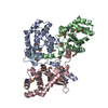
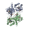
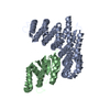
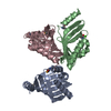
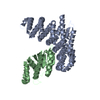
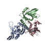
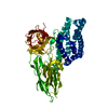
Crystal structure of 5-methylthioribose transporter solute binding protein TLET_1677 from Thermotoga lettingae TMO TARGET EFI-511109 in complex with 5-methylthioribose
ComponentsPeriplasmic binding protein
KeywordsTRANSPORT PROTEIN / SUGAR TRANSPORTER / ENZYME FUNCTION INITIATIVE / EFI / STRUCTURAL GENOMICS
Function / homologyPeriplasmic binding protein / Periplasmic binding protein domain / Response regulator / Periplasmic binding protein-like I / Rossmann fold / 3-Layer(aba) Sandwich / Alpha Beta / 5-S-methyl-5-thio-alpha-D-ribofuranose / Periplasmic binding protein/LacI transcriptional regulator
Function and homology information
Biological speciesThermotoga lettingae TMO (bacteria)
MethodX-RAY DIFFRACTION / SYNCHROTRON / SAD / Resolution: 1.75 Å
AuthorsPatskovsky, Y. / Toro, R. / Bhosle, R. / Al obaidi, N. / Morisco, L.L. / Wasserman, S.R. / Chamala, S. / Attonito, J.D. / Scott glenn, A. / Chowdhury, S. ...Patskovsky, Y. / Toro, R. / Bhosle, R. / Al obaidi, N. / Morisco, L.L. / Wasserman, S.R. / Chamala, S. / Attonito, J.D. / Scott glenn, A. / Chowdhury, S. / Lafleur, J. / Hillerich, B. / Siede, R.D. / Love, J. / Whalen, K.L. / Gerlt, J.A. / Almo, S.C. / Enzyme Function Initiative (EFI)
CitationJournal: To be Published
Title: Crystal structure of 5-methylthioribose binding protein TLET_1677 from Thermotoga lettingae TARGET EFI-511109
Authors: Patskovsky, Y. / Toro, R. / Bhosle, R. / Al obaidi, N. / Chamala, S. / Scott glenn, A. / Attonito, J.D. / Chowdhury, S. / Lafleur, J. / Siedel, R.D. / Morisco, L.L. / Wasserman, S.R. / ...Authors: Patskovsky, Y. / Toro, R. / Bhosle, R. / Al obaidi, N. / Chamala, S. / Scott glenn, A. / Attonito, J.D. / Chowdhury, S. / Lafleur, J. / Siedel, R.D. / Morisco, L.L. / Wasserman, S.R. / Hillerich, B. / Love, J. / Whalen, K.L. / Gerlt, J.A. / Almo, S.C.
History
DepositionDec 13, 2014Deposition site: RCSB / Processing site: RCSB
Revision 1.0Dec 24, 2014Provider: repository / Type: Initial release
Revision 1.1Jan 24, 2018Group: Structure summary / Category: audit_author / Item: _audit_author.name
Revision 2.0Jul 29, 2020Group: Atomic model / Data collection ...Atomic model / Data collection / Database references / Derived calculations / Structure summary
Category: atom_site / chem_comp ...atom_site / chem_comp / entity / pdbx_chem_comp_identifier / pdbx_entity_nonpoly / struct_ref_seq_dif / struct_site / struct_site_gen
Item: _atom_site.auth_atom_id / _atom_site.label_atom_id ..._atom_site.auth_atom_id / _atom_site.label_atom_id / _chem_comp.name / _chem_comp.type / _entity.pdbx_description / _pdbx_entity_nonpoly.name / _struct_ref_seq_dif.details
Description: Carbohydrate remediation / Provider: repository / Type: Remediation
Revision 2.1Feb 28, 2024Group: Data collection / Database references / Structure summary
Category: chem_comp / chem_comp_atom ...chem_comp / chem_comp_atom / chem_comp_bond / database_2
Item: _chem_comp.pdbx_synonyms / _database_2.pdbx_DOI / _database_2.pdbx_database_accession

-
Structure visualization

Structure viewerMolecule:
MolmilJmol/JSmol

Downloads & links

-
Assembly

Deposited unit
A: Periplasmic binding protein
B: Periplasmic binding protein
C: Periplasmic binding protein
D: Periplasmic binding protein
hetero molecules


Theoretical massNumber of molelcules
Total (without water)147,0748
Polymers146,3534
Non-polymers7214
Water8,575476
1
A: Periplasmic binding protein
C: Periplasmic binding protein
hetero molecules


Theoretical massNumber of molelcules
Total (without water)73,5374
Polymers73,1772
Non-polymers3602
Water362
TypeNameSymmetry operationNumber
identity operation1_555x,y,z1
Buried area3000 Å2
ΔGint-23 kcal/mol
Surface area24380 Å2
MethodPISA
2
B: Periplasmic binding protein
D: Periplasmic binding protein
hetero molecules


Theoretical massNumber of molelcules
Total (without water)73,5374
Polymers73,1772
Non-polymers3602
Water362
TypeNameSymmetry operationNumber
identity operation1_555x,y,z1
Buried area2980 Å2
ΔGint-22 kcal/mol
Surface area24530 Å2
MethodPISA
Unit cell
Length a, b, c (Å)57.622, 67.848, 83.653
Angle α, β, γ (deg.)91.83, 92.39, 89.93
Int Tables number1
Space group name H-MP1
Noncrystallographic symmetry (NCS)NCS oper:
IDCodeMatrixVector
1given(1), (1), (1)
2given(0.999996, 0.000352, -0.002893), (0.000376, -0.999965, 0.008405), (-0.00289, -0.008406, -0.99996)0.01663, 65.3016, 21.83962
3given(-0.999996, 0.000311, 0.002959), (-0.000344, -0.999941, -0.010893), (0.002955, -0.010894, 0.999936)97.93922, 65.5398, 0.27788
4given(-1, 0.000197, -0.000738), (0.000197, 1, -0.000356), (0.000738, -0.000356, -1)97.90417, -0.02871, 21.37169

-
Components

#1: Protein
Periplasmic binding protein


Mass: 36588.270 Da / Num. of mol.: 4 / Fragment: UNP RESIDUES 22-348
Source method: isolated from a genetically manipulated source
Source: (gene. exp.) Thermotoga lettingae TMO (bacteria) / Gene: Tlet_1677 / Plasmid: PET / Production host: Escherichia coli (E. coli) / Strain (production host): BL21(DE3) / References: UniProt: A8F7U7
#2: Sugar
ChemComp-SR1 / 5-S-methyl-5-thio-alpha-D-ribofuranose / 5-S-methyl-5-thio-alpha-D-ribose / 5-S-methyl-5-thio-D-ribose / 5-S-methyl-5-thio-ribose


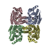
Type: D-saccharide, alpha linking / Mass: 180.222 Da / Num. of mol.: 4 / Source method: obtained synthetically / Formula: C6H12O4S
IdentifierTypeProgram
a-D-Ribf5SMeIUPAC CARBOHYDRATE SYMBOLPDB-CARE 1.0
#3: Water ChemComp-HOH / water


Mass: 18.015 Da / Num. of mol.: 476 / Source method: isolated from a natural source / Formula: H2O

-
Experimental details

-
Experiment

ExperimentMethod: X-RAY DIFFRACTION / Number of used crystals: 1

-
Sample preparation

CrystalDensity Matthews: 2.23 Å3/Da / Density % sol: 44.88 %
Crystal growTemperature: 294 K / Method: vapor diffusion, sitting drop / pH: 7.5
Details: PROTEIN (10 MM HEPES PH 7.5, 5 MM DTT); RESERVOIR: 0.1 M HEPES-NAOH, PH 7.5, 0.2 M MAGNESIUM CHLORIDE, 30% PPG P400; CRYOPROTECTION: RESERVOIR SOLUTION; VAPOR DIFFUSION, SITTING DROP, TEMPERATURE 294K;

-
Data collection

DiffractionMean temperature: 100 K
Diffraction sourceSource: SYNCHROTRON / Site: APS / Beamline: 31-ID / Wavelength: 0.9793
DetectorType: RAYONIX MX225HE / Detector: CCD / Date: Nov 22, 2014 / Details: MIRRORS
RadiationMonochromator: GRAPHITE / Protocol: SINGLE WAVELENGTH / Monochromatic (M) / Laue (L): M / Scattering type: x-ray
Radiation wavelengthWavelength: 0.9793 Å / Relative weight: 1
ReflectionResolution: 1.75→50 Å / Num. obs: 128230 / % possible obs: 97.1 % / Observed criterion σ(I): -3 / Redundancy: 3.9 % / Rmerge(I) obs: 0.137 / Net I/σ(I): 10
Reflection shellResolution: 1.75→1.78 Å / Redundancy: 3.6 % / Rmerge(I) obs: 0.71 / Mean I/σ(I) obs: 2.1 / % possible all: 94.9

-
Processing

Software
NameVersionClassification
SHELXmodel building
ARP/wARPmodel building
REFMAC5.8.0073refinement
HKL-3000data reduction
HKL-3000data scaling
SHELXphasing
RefinementMethod to determine structure: SAD / Resolution: 1.75→50 Å / Cor.coef. Fo:Fc: 0.953 / Cor.coef. Fo:Fc free: 0.928 / SU B: 12.196 / SU ML: 0.173 / Cross valid method: THROUGHOUT / ESU R: 0.14 / ESU R Free: 0.139 / Stereochemistry target values: MAXIMUM LIKELIHOOD / Details: HYDROGENS HAVE BEEN ADDED IN THE RIDING POSITIONS
RfactorNum. reflection% reflectionSelection details
Rfree0.26356 3771 3 %RANDOM
Rwork0.21444 ---
obs0.21593 119981 96.95 %-
Solvent computationIon probe radii: 0.8 Å / Shrinkage radii: 0.8 Å / VDW probe radii: 1.2 Å / Solvent model: MASK
Displacement parametersBiso mean: 33.26 Å2
Baniso -1Baniso -2Baniso -3
1--3.67 Å2-0.31 Å2-0.41 Å2
2---3.8 Å20.37 Å2
3---7.44 Å2
Refinement stepCycle: LAST / Resolution: 1.75→50 Å
ProteinNucleic acidLigandSolventTotal
Num. atoms9868 0 44 476 10388
Refine LS restraints
Refine-IDTypeDev idealDev ideal targetNumber
X-RAY DIFFRACTIONr_bond_refined_d0.0130.01910127
X-RAY DIFFRACTIONr_bond_other_d0.0010.029979
X-RAY DIFFRACTIONr_angle_refined_deg1.6011.96613733
X-RAY DIFFRACTIONr_angle_other_deg0.835322888
X-RAY DIFFRACTIONr_dihedral_angle_1_deg6.36851276
X-RAY DIFFRACTIONr_dihedral_angle_2_deg36.48424.695443
X-RAY DIFFRACTIONr_dihedral_angle_3_deg14.933151776
X-RAY DIFFRACTIONr_dihedral_angle_4_deg20.3691568
X-RAY DIFFRACTIONr_chiral_restr0.0910.21592
X-RAY DIFFRACTIONr_gen_planes_refined0.0070.02111397
X-RAY DIFFRACTIONr_gen_planes_other0.0010.022191
X-RAY DIFFRACTIONr_nbd_refined
X-RAY DIFFRACTIONr_nbd_other
X-RAY DIFFRACTIONr_nbtor_refined
X-RAY DIFFRACTIONr_nbtor_other
X-RAY DIFFRACTIONr_xyhbond_nbd_refined
X-RAY DIFFRACTIONr_xyhbond_nbd_other
X-RAY DIFFRACTIONr_metal_ion_refined
X-RAY DIFFRACTIONr_metal_ion_other
X-RAY DIFFRACTIONr_symmetry_vdw_refined
X-RAY DIFFRACTIONr_symmetry_vdw_other
X-RAY DIFFRACTIONr_symmetry_hbond_refined
X-RAY DIFFRACTIONr_symmetry_hbond_other
X-RAY DIFFRACTIONr_symmetry_metal_ion_refined
X-RAY DIFFRACTIONr_symmetry_metal_ion_other
X-RAY DIFFRACTIONr_mcbond_it5.6721.5225104
X-RAY DIFFRACTIONr_mcbond_other5.6651.5225103
X-RAY DIFFRACTIONr_mcangle_it6.1822.2216371
X-RAY DIFFRACTIONr_mcangle_other6.1842.2216372
X-RAY DIFFRACTIONr_scbond_it10.8562.4885023
X-RAY DIFFRACTIONr_scbond_other10.8552.4885024
X-RAY DIFFRACTIONr_scangle_it
X-RAY DIFFRACTIONr_scangle_other11.1813.2467360
X-RAY DIFFRACTIONr_long_range_B_refined11.6425.56411857
X-RAY DIFFRACTIONr_long_range_B_other11.6425.56411858
X-RAY DIFFRACTIONr_rigid_bond_restr
X-RAY DIFFRACTIONr_sphericity_free
X-RAY DIFFRACTIONr_sphericity_bonded
LS refinement shellResolution: 1.75→1.795 Å / Total num. of bins used: 20
RfactorNum. reflection% reflection
Rfree0.345 291 -
Rwork0.335 8540 -
obs--93.73 %
Refinement TLS params.

Method: refined / Refine-ID: X-RAY DIFFRACTION

IDL112)L122)L132)L222)L232)L332)S11 (Å °)S12 (Å °)S13 (Å °)S21 (Å °)S22 (Å °)S23 (Å °)S31 (Å °)S32 (Å °)S33 (Å °)T112)T122)T132)T222)T232)T332)Origin x (Å)Origin y (Å)Origin z (Å)
12.51880.0897-0.21011.7457-0.10160.4295-0.0096-0.1745-0.23150.19760.00730.00880.0346-0.02490.00220.26530.02240.01950.23410.03880.025248.995815.766228.1799
22.4017-0.42060.2551.6949-0.1380.4197-0.01290.18950.2358-0.17960.00020.0092-0.0276-0.01830.01270.2694-0.00980.02660.23720.04560.032448.290349.7741-7.1626
32.16310.37770.2751.60650.16990.4329-0.008-0.18460.21580.1688-0.0033-0.0321-0.03490.01640.01130.26450.02570.02560.23940.00780.032349.594949.629528.5266
42.2903-0.1055-0.11951.90080.22590.4334-0.00120.1888-0.2433-0.21630.0181-0.08530.02070.0387-0.01690.2708-0.0040.02590.24570.00520.036549.4815.5388-7.0396
Refinement TLS group
IDRefine-IDRefine TLS-IDAuth asym-IDAuth seq-ID
1X-RAY DIFFRACTION1A13 - 327
2X-RAY DIFFRACTION2B7 - 327
3X-RAY DIFFRACTION3C5 - 327
4X-RAY DIFFRACTION4D7 - 327

+
About Yorodumi

-
News

-
Feb 9, 2022. New format data for meta-information of EMDB entries

New format data for meta-information of EMDB entries

  • Version 3 of the EMDB header file is now the official format.
  • The previous official version 1.9 will be removed from the archive.

Related info.:EMDB header

External links:wwPDB to switch to version 3 of the EMDB data model

-
Aug 12, 2020. Covid-19 info

Covid-19 info

URL: https://pdbjlvh1.pdbj.org/emnavi/covid19.php

New page: Covid-19 featured information page in EM Navigator.

Related info.:Covid-19 info / Mar 5, 2020. Novel coronavirus structure data

+
Mar 5, 2020. Novel coronavirus structure data

Novel coronavirus structure data

Related info.:Yorodumi Speices / Aug 12, 2020. Covid-19 info

External links:COVID-19 featured content - PDBj / Molecule of the Month (242):Coronavirus Proteases

+
Jan 31, 2019. EMDB accession codes are about to change! (news from PDBe EMDB page)

EMDB accession codes are about to change! (news from PDBe EMDB page)

  • The allocation of 4 digits for EMDB accession codes will soon come to an end. Whilst these codes will remain in use, new EMDB accession codes will include an additional digit and will expand incrementally as the available range of codes is exhausted. The current 4-digit format prefixed with “EMD-” (i.e. EMD-XXXX) will advance to a 5-digit format (i.e. EMD-XXXXX), and so on. It is currently estimated that the 4-digit codes will be depleted around Spring 2019, at which point the 5-digit format will come into force.
  • The EM Navigator/Yorodumi systems omit the EMD- prefix.

Related info.:Q: What is EMD? / ID/Accession-code notation in Yorodumi/EM Navigator

External links:EMDB Accession Codes are Changing Soon! / Contact to PDBj

+
Jul 12, 2017. Major update of PDB

Major update of PDB

  • wwPDB released updated PDB data conforming to the new PDBx/mmCIF dictionary.
  • This is a major update changing the version number from 4 to 5, and with Remediation, in which all the entries are updated.
  • In this update, many items about electron microscopy experimental information are reorganized (e.g. em_software).
  • Now, EM Navigator and Yorodumi are based on the updated data.

External links:wwPDB Remediation / Enriched Model Files Conforming to OneDep Data Standards Now Available in the PDB FTP Archive

-
Yorodumi

Thousand views of thousand structures

  • Yorodumi is a browser for structure data from EMDB, PDB, SASBDB, etc.
  • This page is also the successor to EM Navigator detail page, and also detail information page/front-end page for Omokage search.
  • The word "yorodu" (or yorozu) is an old Japanese word meaning "ten thousand". "mi" (miru) is to see.

Related info.:EMDB / PDB / SASBDB / Comparison of 3 databanks / Yorodumi Search / Aug 31, 2016. New EM Navigator & Yorodumi / Yorodumi Papers / Jmol/JSmol / Function and homology information / Changes in new EM Navigator and Yorodumi

Read more