 Movie
Movie Controller

[English] 日本語
 Yorodumi
- PDB-2y3w: N-terminal head domain and beginning of coiled coil domain of Dan... -
+ Open data

- Basic information
| Entry | Database: PDB / ID: 2y3w | ||||||
|---|---|---|---|---|---|---|---|
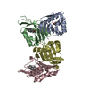
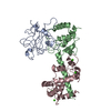
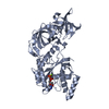
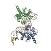
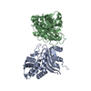
| Title | N-terminal head domain and beginning of coiled coil domain of Danio rerio SAS-6 | ||||||
 Components Components | SPINDLE ASSEMBLY ABNORMAL PROTEIN 6 HOMOLOG | ||||||
 Keywords Keywords | STRUCTURAL PROTEIN / CYTOSKELETON / BASAL BODY / CENTRIOLE / CARTWHEEL / CARTWHEEL HUB | ||||||
| Function / homology |  Function and homology information Function and homology informationpositive regulation of mitotic spindle organization / deuterosome / positive regulation of centriole replication / nuclear division / embryonic cleavage / positive regulation of spindle assembly / centriole replication / centrosome duplication / positive regulation of G1/S transition of mitotic cell cycle / centriole ...positive regulation of mitotic spindle organization / deuterosome / positive regulation of centriole replication / nuclear division / embryonic cleavage / positive regulation of spindle assembly / centriole replication / centrosome duplication / positive regulation of G1/S transition of mitotic cell cycle / centriole / mitotic spindle organization / spermatogenesis / centrosome / cytoplasm Similarity search - Function | ||||||
| Biological species |  | ||||||
| Method |  X-RAY DIFFRACTION / SYNCHROTRON / MOLECULAR REPLACEMENT / Resolution: 1.98 Å X-RAY DIFFRACTION / SYNCHROTRON / MOLECULAR REPLACEMENT / Resolution: 1.98 Å | ||||||
 Authors Authors | van Breugel, M. | ||||||
 Citation Citation | Journal: Science / Year: 2011 Title: Structures of SAS-6 suggest its organization in centrioles. Authors: van Breugel, M. / Hirono, M. / Andreeva, A. / Yanagisawa, H.A. / Yamaguchi, S. / Nakazawa, Y. / Morgner, N. / Petrovich, M. / Ebong, I.O. / Robinson, C.V. / Johnson, C.M. / Veprintsev, D. / Zuber, B. | ||||||
| History |
| ||||||
| Remark 700 | SHEET DETERMINATION METHOD: DSSP THE SHEETS PRESENTED AS "AA" IN EACH CHAIN ON SHEET RECORDS BELOW ... SHEET DETERMINATION METHOD: DSSP THE SHEETS PRESENTED AS "AA" IN EACH CHAIN ON SHEET RECORDS BELOW IS ACTUALLY AN 7-STRANDED BARREL THIS IS REPRESENTED BY A 8-STRANDED SHEET IN WHICH THE FIRST AND LAST STRANDS ARE IDENTICAL. THE SHEETS PRESENTED AS "BA" IN EACH CHAIN ON SHEET RECORDS BELOW IS ACTUALLY AN 7-STRANDED BARREL THIS IS REPRESENTED BY A 8-STRANDED SHEET IN WHICH THE FIRST AND LAST STRANDS ARE IDENTICAL. THE SHEETS PRESENTED AS "CA" IN EACH CHAIN ON SHEET RECORDS BELOW IS ACTUALLY AN 7-STRANDED BARREL THIS IS REPRESENTED BY A 8-STRANDED SHEET IN WHICH THE FIRST AND LAST STRANDS ARE IDENTICAL. |
- Structure visualization
| Structure viewer | Molecule: MolmilJmol/JSmol |
|---|
- Downloads & links
-Download
| PDBx/mmCIF format | 2y3w.cif.gz | 105.5 KB | Display | PDBx/mmCIF format |
|---|---|---|---|---|
| PDB format | pdb2y3w.ent.gz | 81.3 KB | Display | PDB format |
| PDBx/mmJSON format | 2y3w.json.gz | Tree view | PDBx/mmJSON format | |
| Others |  Other downloads Other downloads |
-Validation report
| Arichive directory | https://data.pdbj.org/pub/pdb/validation_reports/y3/2y3wftp://data.pdbj.org/pub/pdb/validation_reports/y3/2y3w | HTTPS FTP |
|---|
-Related structure data
| Related structure data |  2y3vSC S: Starting model for refinement C: citing same article ( |
|---|---|
| Similar structure data |


-Links
 PDBj
PDBj- Assembly
| Deposited unit | 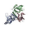
| ||||||||
|---|---|---|---|---|---|---|---|---|---|
| 1 | 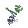
| ||||||||
| 2 | 
| ||||||||
| Unit cell |
| ||||||||
| Noncrystallographic symmetry (NCS) | NCS oper: (Code: given Matrix: (-0.9826, -0.1455, 0.1153), Vector: |
-Components
| #1: Protein | Mass: 20987.885 Da / Num. of mol.: 3 Fragment: HEAD DOMAIN AND START OF COILED-COIL DOMAIN, RESIDUES 1-179 Mutation: YES Source method: isolated from a genetically manipulated source Source: (gene. exp.)  #2: Water | ChemComp-HOH / |  Mass: 18.015 Da / Num. of mol.: 146 / Source method: isolated from a natural source / Formula: H2O Mass: 18.015 Da / Num. of mol.: 146 / Source method: isolated from a natural source / Formula: H2OCompound details | ENGINEERED RESIDUE IN CHAIN A, PHE 131 TO ASP ENGINEERED RESIDUE IN CHAIN B, PHE 131 TO ASP ...ENGINEERED | Sequence details | THE N-TERMINAL THREE AMINO ACIDS (GPH) STEM FROM THE EXPRESSION VECTOR. THE F131D MUTATION WAS ...THE N-TERMINAL THREE AMINO ACIDS (GPH) STEM FROM THE EXPRESSION | |
|---|
-Experimental details
-Experiment
| Experiment | Method: X-RAY DIFFRACTION / Number of used crystals: 1 |
|---|
- Sample preparation
| Crystal | Density Matthews: 2.39 Å3/Da / Density % sol: 48.54 % / Description: NONE |
|---|---|
| Crystal grow | pH: 9.2 / Details: 0.1 M CHES PH 9.2, 36% PEG 600, 1MM DTT. |
-Data collection
| Diffraction | Mean temperature: 77 K |
|---|---|
| Diffraction source | Source: SYNCHROTRON / Site: ESRF  / Beamline: BM14 / Wavelength: 0.9184 / Beamline: BM14 / Wavelength: 0.9184 |
| Detector | Type: MARMOSAIC 225 mm CCD / Detector: CCD / Date: May 1, 2010 |
| Radiation | Protocol: SINGLE WAVELENGTH / Monochromatic (M) / Laue (L): M / Scattering type: x-ray |
| Radiation wavelength | Wavelength: 0.9184 Å / Relative weight: 1 |
| Reflection | Resolution: 1.98→35.49 Å / Num. obs: 38948 / % possible obs: 99.9 % / Observed criterion σ(I): 0 / Redundancy: 11.5 % / Rmerge(I) obs: 0.09 / Net I/σ(I): 12.5 |
| Reflection shell | Resolution: 1.98→2.09 Å / Redundancy: 11.4 % / Rmerge(I) obs: 1.5 / Mean I/σ(I) obs: 1.6 / % possible all: 100 |
- Processing
| Software |
| |||||||||||||||||||||||||||||||||||||||||||||||||||||||||||||||||||||||||||||||||||||||||||||||||||||||||
|---|---|---|---|---|---|---|---|---|---|---|---|---|---|---|---|---|---|---|---|---|---|---|---|---|---|---|---|---|---|---|---|---|---|---|---|---|---|---|---|---|---|---|---|---|---|---|---|---|---|---|---|---|---|---|---|---|---|---|---|---|---|---|---|---|---|---|---|---|---|---|---|---|---|---|---|---|---|---|---|---|---|---|---|---|---|---|---|---|---|---|---|---|---|---|---|---|---|---|---|---|---|---|---|---|---|---|
| Refinement | Method to determine structure: MOLECULAR REPLACEMENT Starting model: PDB ENTRY 2Y3V Resolution: 1.98→35.492 Å / SU ML: 0.32 / σ(F): 2.05 / Phase error: 26.47 / Stereochemistry target values: ML Details: RESIDUES A-2, A36 TO A41, AND A177 TO A179, RESIDUES B-2 TO B0, B16 TO B20, B112 TO B117, B178 TO B179, RESIDUES C-2 TO C1, C15 TO C21, C34 TO C43, C63 TO C64, C73 TO C78, C112 TO C118, C129 ...Details: RESIDUES A-2, A36 TO A41, AND A177 TO A179, RESIDUES B-2 TO B0, B16 TO B20, B112 TO B117, B178 TO B179, RESIDUES C-2 TO C1, C15 TO C21, C34 TO C43, C63 TO C64, C73 TO C78, C112 TO C118, C129 TO C136, AND C145 TO C179 ARE DISORDERED. CHAIN C SHOWED GOOD DENSITY MAINLY IN THE REGIONS CONTACTING CHAIN A AND B.
| |||||||||||||||||||||||||||||||||||||||||||||||||||||||||||||||||||||||||||||||||||||||||||||||||||||||||
| Solvent computation | Shrinkage radii: 0.61 Å / VDW probe radii: 0.9 Å / Solvent model: FLAT BULK SOLVENT MODEL / Bsol: 60 Å2 / ksol: 0.352 e/Å3 | |||||||||||||||||||||||||||||||||||||||||||||||||||||||||||||||||||||||||||||||||||||||||||||||||||||||||
| Displacement parameters | Biso mean: 58.72 Å2
| |||||||||||||||||||||||||||||||||||||||||||||||||||||||||||||||||||||||||||||||||||||||||||||||||||||||||
| Refinement step | Cycle: LAST / Resolution: 1.98→35.492 Å
| |||||||||||||||||||||||||||||||||||||||||||||||||||||||||||||||||||||||||||||||||||||||||||||||||||||||||
| Refine LS restraints |
| |||||||||||||||||||||||||||||||||||||||||||||||||||||||||||||||||||||||||||||||||||||||||||||||||||||||||
| LS refinement shell |
|
