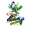
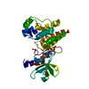
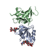
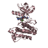
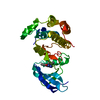
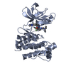
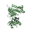
Entry Database : PDB / ID : 2og8Title crystal structure of aminoquinazoline 36 bound to Lck Proto-oncogene tyrosine-protein kinase LCK Keywords / / Function / homology Function Domain/homology Component
/ / / / / / / / / / / / / / / / / / / / / / / / / / / / / / / / / / / / / / / / / / / / / / / / / / / / / / / / / / / / / / / / / / / / / / / / / / / / / / / / / / / / / / / / / / / / / / / / / / / / / / / / / / / / / / / / / / / / / / / / / / Biological species Homo sapiens (human)Method / / Resolution : 2.3 Å Authors Huang, X. Journal : J.Med.Chem. / Year : 2006Title : Discovery of Aminoquinazolines as Potent, Orally Bioavailable Inhibitors of Lck: Synthesis, SAR, and in Vivo Anti-Inflammatory ActivityAuthors: DiMauro, E.F. / Newcomb, J. / Nunes, J.J. / Bemis, J.E. / Boucher, C. / Buchanan, J.L. / Buckner, W.H. / Cee, V.J. / Chai, L. / Deak, H.L. / Epstein, L.F. / Faust, T. / Gallant, P. / Geuns- ... Authors : DiMauro, E.F. / Newcomb, J. / Nunes, J.J. / Bemis, J.E. / Boucher, C. / Buchanan, J.L. / Buckner, W.H. / Cee, V.J. / Chai, L. / Deak, H.L. / Epstein, L.F. / Faust, T. / Gallant, P. / Geuns-Meyer, S.D. / Gore, A. / Gu, Y. / Henkle, B. / Hodous, B.L. / Hsieh, F. / Huang, X. / Kim, J.L. / Lee, J.H. / Martin, M.W. / Masse, C.E. / McGowan, D.C. / Metz, D. / Morgenstern, K.A. / Oliveira-dos-Santos, A. / Patel, V.F. / Powers, D. / Rose, P.E. / Schneider, S. / Tomlinson, S.A. / Tudor, Y.Y. / Turci, S.M. / Welcher, A.A. / White, R.D. / Zhao, H. / Zhu, L. / Zhu, X. History Deposition Jan 5, 2007 Deposition site / Processing site Revision 1.0 Feb 27, 2007 Provider / Type Revision 1.1 May 1, 2008 Group Revision 1.2 Jul 13, 2011 Group Revision 1.3 Aug 30, 2023 Group / Database references / Refinement descriptionCategory chem_comp_atom / chem_comp_bond ... chem_comp_atom / chem_comp_bond / database_2 / pdbx_initial_refinement_model / struct_ref_seq_dif Item / _database_2.pdbx_database_accession / _struct_ref_seq_dif.details
Show all Show less
 Movie
Movie Controller


 Open data
 Basic information
 Components
 Keywords
 Function and homology information
 Homo sapiens (human)
 X-RAY DIFFRACTION /
  Authors
 Citation
 Structure visualization
 Downloads & links
 Other downloads
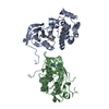


 PDBj
PDBj


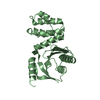
 Assembly
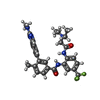
 Mass: 564.601 Da / Num. of mol.: 2 / Source method: obtained synthetically / Formula: C30H31F3N6O2
 Mass: 18.015 Da / Num. of mol.: 209 / Source method: isolated from a natural source / Formula: H2O
 Sample preparation
 Processing