 Movie
Movie Controller


+ Open data

- Basic information
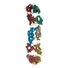
| Entry | Database: PDB / ID: 4rsi | ||||||
|---|---|---|---|---|---|---|---|
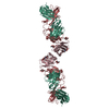
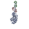
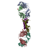
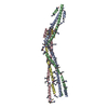
| Title | Yeast Smc2-Smc4 hinge domain with extended coiled coils | ||||||
 Components Components |
| ||||||
 Keywords Keywords | CELL CYCLE / Smc hinge domain with coiled coil / chromosomal condensation | ||||||
| Function / homology |  Function and homology information Function and homology informationnegative regulation of meiotic DNA double-strand break formation / tRNA gene clustering / Condensation of Prometaphase Chromosomes / condensin complex / DNA secondary structure binding / rDNA chromatin condensation / mitotic chromosome condensation / chromosome condensation / minor groove of adenine-thymine-rich DNA binding / mitotic sister chromatid segregation ...negative regulation of meiotic DNA double-strand break formation / tRNA gene clustering / Condensation of Prometaphase Chromosomes / condensin complex / DNA secondary structure binding / rDNA chromatin condensation / mitotic chromosome condensation / chromosome condensation / minor groove of adenine-thymine-rich DNA binding / mitotic sister chromatid segregation / condensed chromosome / double-stranded DNA binding / cell division / chromatin binding / chromatin / ATP hydrolysis activity / mitochondrion / ATP binding / nucleus / cytoplasm Similarity search - Function | ||||||
| Biological species |  | ||||||
| Method |  X-RAY DIFFRACTION / SYNCHROTRON / SAD / Resolution: 2.9 Å X-RAY DIFFRACTION / SYNCHROTRON / SAD / Resolution: 2.9 Å | ||||||
 Authors Authors | Soh, Y.M. / Shin, H.C. / Oh, B.H. | ||||||
 Citation Citation | Journal: Mol.Cell / Year: 2015 Title: Molecular Basis for SMC Rod Formation and Its Dissolution upon DNA Binding. Authors: Soh, Y.M. / Burmann, F. / Shin, H.C. / Oda, T. / Jin, K.S. / Toseland, C.P. / Kim, C. / Lee, H. / Kim, S.J. / Kong, M.S. / Durand-Diebold, M.L. / Kim, Y.G. / Kim, H.M. / Lee, N.K. / Sato, M. ...Authors: Soh, Y.M. / Burmann, F. / Shin, H.C. / Oda, T. / Jin, K.S. / Toseland, C.P. / Kim, C. / Lee, H. / Kim, S.J. / Kong, M.S. / Durand-Diebold, M.L. / Kim, Y.G. / Kim, H.M. / Lee, N.K. / Sato, M. / Oh, B.H. / Gruber, S. | ||||||
| History |
|
- Structure visualization
| Structure viewer | Molecule: MolmilJmol/JSmol |
|---|
- Downloads & links
-Download
| PDBx/mmCIF format | 4rsi.cif.gz | 146.8 KB | Display | PDBx/mmCIF format |
|---|---|---|---|---|
| PDB format | pdb4rsi.ent.gz | 112.1 KB | Display | PDB format |
| PDBx/mmJSON format | 4rsi.json.gz | Tree view | PDBx/mmJSON format | |
| Others |  Other downloads Other downloads |
-Validation report
| Arichive directory | https://data.pdbj.org/pub/pdb/validation_reports/rs/4rsiftp://data.pdbj.org/pub/pdb/validation_reports/rs/4rsi | HTTPS FTP |
|---|
-Related structure data


-Links
 PDBj
PDBj- Assembly
| Deposited unit | 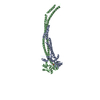
| ||||||||
|---|---|---|---|---|---|---|---|---|---|
| 1 |
| ||||||||
| Unit cell |
|
-Components
| #1: Protein | Mass: 45382.734 Da / Num. of mol.: 1 / Fragment: Smc2 hinge Source method: isolated from a genetically manipulated source Details: separate expression co-purification Source: (gene. exp.) Strain: ATCC 204508 / S288c / Gene: Saccharomyces cerevisiae, SMC2, YFR031C / Plasmid: pProExHTa / Production host:  | ||
|---|---|---|---|
| #2: Protein | Mass: 45231.340 Da / Num. of mol.: 1 / Fragment: Smc4 hinge Source method: isolated from a genetically manipulated source Details: stop codon, culture in minimal media, co-purification Source: (gene. exp.) Strain: ATCC 204508 / S288c / Gene: L9449.5, Saccharomyces cerevisiae, SMC4, YLR086W / Plasmid: pET30a / Production host: | ||
| #3: Chemical |   Mass: 94.971 Da / Num. of mol.: 2 / Source method: obtained synthetically / Formula: PO4 Mass: 94.971 Da / Num. of mol.: 2 / Source method: obtained synthetically / Formula: PO4#4: Water | ChemComp-HOH / |  Mass: 18.015 Da / Num. of mol.: 8 / Source method: isolated from a natural source / Formula: H2O Mass: 18.015 Da / Num. of mol.: 8 / Source method: isolated from a natural source / Formula: H2O |
-Experimental details
-Experiment
| Experiment | Method: X-RAY DIFFRACTION / Number of used crystals: 1 |
|---|
- Sample preparation
| Crystal | Density Matthews: 3.92 Å3/Da / Density % sol: 68.59 % |
|---|---|
| Crystal grow | Temperature: 293.15 K / Method: vapor diffusion, hanging drop / pH: 6 Details: 16% PEG 300, 0.1 M Na/K phosphate pH 6.0, 8% glycerol, 10 mM dithiothreitol (DTT), VAPOR DIFFUSION, HANGING DROP, temperature 293.15K |
-Data collection
| Diffraction | Mean temperature: 110 K |
|---|---|
| Diffraction source | Source: SYNCHROTRON / Site: Photon Factory  / Beamline: BL-17A / Wavelength: 0.9789 Å / Beamline: BL-17A / Wavelength: 0.9789 Å |
| Detector | Type: ADSC QUANTUM 270 / Detector: CCD / Date: Nov 8, 2012 |
| Radiation | Monochromator: Numerical link type double crystal monochromator, liquid nitrogen cooling Si(111) Protocol: SINGLE WAVELENGTH / Monochromatic (M) / Laue (L): M / Scattering type: x-ray |
| Radiation wavelength | Wavelength: 0.9789 Å / Relative weight: 1 |
| Reflection | Resolution: 2.9→50 Å / Num. all: 49139 / Num. obs: 28485 / % possible obs: 90.2 % / Observed criterion σ(F): 2.5 / Observed criterion σ(I): 2.5 / Redundancy: 4.4 % / Rsym value: 0.09 / Net I/σ(I): 33.5 |
- Processing
| Software |
| |||||||||||||||||||||||||||||||||||||||||||||||||||||||||||||||||||||||||||||||||||||||||||||||||||||||||||||||||||||||||||||||||||||||||||||||||||
|---|---|---|---|---|---|---|---|---|---|---|---|---|---|---|---|---|---|---|---|---|---|---|---|---|---|---|---|---|---|---|---|---|---|---|---|---|---|---|---|---|---|---|---|---|---|---|---|---|---|---|---|---|---|---|---|---|---|---|---|---|---|---|---|---|---|---|---|---|---|---|---|---|---|---|---|---|---|---|---|---|---|---|---|---|---|---|---|---|---|---|---|---|---|---|---|---|---|---|---|---|---|---|---|---|---|---|---|---|---|---|---|---|---|---|---|---|---|---|---|---|---|---|---|---|---|---|---|---|---|---|---|---|---|---|---|---|---|---|---|---|---|---|---|---|---|---|---|---|
| Refinement | Method to determine structure: SAD / Resolution: 2.9→38.922 Å / SU ML: 0.44 / σ(F): 1.85 / Phase error: 28.12 / Stereochemistry target values: ML
| |||||||||||||||||||||||||||||||||||||||||||||||||||||||||||||||||||||||||||||||||||||||||||||||||||||||||||||||||||||||||||||||||||||||||||||||||||
| Solvent computation | Shrinkage radii: 0.9 Å / VDW probe radii: 1.11 Å / Solvent model: FLAT BULK SOLVENT MODEL | |||||||||||||||||||||||||||||||||||||||||||||||||||||||||||||||||||||||||||||||||||||||||||||||||||||||||||||||||||||||||||||||||||||||||||||||||||
| Refinement step | Cycle: LAST / Resolution: 2.9→38.922 Å
| |||||||||||||||||||||||||||||||||||||||||||||||||||||||||||||||||||||||||||||||||||||||||||||||||||||||||||||||||||||||||||||||||||||||||||||||||||
| Refine LS restraints |
| |||||||||||||||||||||||||||||||||||||||||||||||||||||||||||||||||||||||||||||||||||||||||||||||||||||||||||||||||||||||||||||||||||||||||||||||||||
| LS refinement shell |
|
