- PDB-4rke: Drosophila melanogaster Rab2 bound to GMPPNP -
+
Open data
ID or keywords:
Loading...
-
Basic information
Entry
Database: PDB / ID: 4rke
Title
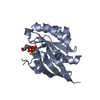
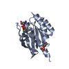
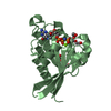
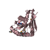
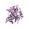
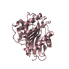
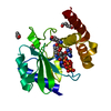
Drosophila melanogaster Rab2 bound to GMPPNP
Components
GH01619p
Keywords
HYDROLASE / GTP hydrolysis
Function / homology
Function and homology information
type II terminal bouton / regulation of glutamate receptor clustering / secretory granule-lysosome fusion / Golgi Cisternae Pericentriolar Stack Reorganization / eye pigment granule organization / RAB geranylgeranylation / postsynapse of neuromuscular junction / T-tubule organization / Rab protein signal transduction / autophagosome-lysosome fusion ...type II terminal bouton / regulation of glutamate receptor clustering / secretory granule-lysosome fusion / Golgi Cisternae Pericentriolar Stack Reorganization / eye pigment granule organization / RAB geranylgeranylation / postsynapse of neuromuscular junction / T-tubule organization / Rab protein signal transduction / autophagosome-lysosome fusion / autolysosome membrane / regulation of postsynapse organization / presynaptic active zone / autophagosome membrane / endoplasmic reticulum exit site / protein secretion / endoplasmic reticulum to Golgi vesicle-mediated transport / vesicle-mediated transport / small monomeric GTPase / cytoplasmic vesicle / vesicle / perikaryon / Golgi membrane / neuronal cell body / GTPase activity / symbiont entry into host cell / GTP binding / magnesium ion binding / Golgi apparatus / plasma membrane Similarity search - Function
: / Small GTPase Rab domain profile. / Small GTPase Rho domain profile. / Small GTPase Ras domain profile. / Ran (Ras-related nuclear proteins) /TC4 subfamily of small GTPases / Rho (Ras homology) subfamily of Ras-like small GTPases / Ras subfamily of RAS small GTPases / Small GTPase / Ras family / Rab subfamily of small GTPases ...: / Small GTPase Rab domain profile. / Small GTPase Rho domain profile. / Small GTPase Ras domain profile. / Ran (Ras-related nuclear proteins) /TC4 subfamily of small GTPases / Rho (Ras homology) subfamily of Ras-like small GTPases / Ras subfamily of RAS small GTPases / Small GTPase / Ras family / Rab subfamily of small GTPases / Small GTP-binding protein domain / P-loop containing nucleotide triphosphate hydrolases / Rossmann fold / P-loop containing nucleoside triphosphate hydrolase / 3-Layer(aba) Sandwich / Alpha Beta Similarity search - Domain/homology
In the structure databanks used in Yorodumi, some data are registered as the other names, "COVID-19 virus" and "2019-nCoV". Here are the details of the virus and the list of structure data.
Jan 31, 2019. EMDB accession codes are about to change! (news from PDBe EMDB page)
EMDB accession codes are about to change! (news from PDBe EMDB page)
The allocation of 4 digits for EMDB accession codes will soon come to an end. Whilst these codes will remain in use, new EMDB accession codes will include an additional digit and will expand incrementally as the available range of codes is exhausted. The current 4-digit format prefixed with “EMD-” (i.e. EMD-XXXX) will advance to a 5-digit format (i.e. EMD-XXXXX), and so on. It is currently estimated that the 4-digit codes will be depleted around Spring 2019, at which point the 5-digit format will come into force.
The EM Navigator/Yorodumi systems omit the EMD- prefix.
Related info.:Q: What is EMD? / ID/Accession-code notation in Yorodumi/EM Navigator
Yorodumi is a browser for structure data from EMDB, PDB, SASBDB, etc.
This page is also the successor to EM Navigator detail page, and also detail information page/front-end page for Omokage search.
The word "yorodu" (or yorozu) is an old Japanese word meaning "ten thousand". "mi" (miru) is to see.
Related info.:EMDB / PDB / SASBDB / Comparison of 3 databanks / Yorodumi Search / Aug 31, 2016. New EM Navigator & Yorodumi / Yorodumi Papers / Jmol/JSmol / Function and homology information / Changes in new EM Navigator and Yorodumi
 Movie
Movie Controller


 Open data
 Basic information
 Components
 Keywords
 Function and homology information
 X-RAY DIFFRACTION /
  Authors
 Citation
 Structure visualization
 Downloads & links
 Other downloads


 PDBj
PDBj


 Assembly
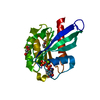


 Mass: 522.196 Da / Num. of mol.: 1 / Source method: obtained synthetically / Formula: C10H17N6O13P3
 Mass: 24.305 Da / Num. of mol.: 1 / Source method: obtained synthetically / Formula: Mg
 Mass: 238.278 Da / Num. of mol.: 2 / Source method: obtained synthetically / Formula: C10H22O6 / Comment: precipitant*YM
 Mass: 18.015 Da / Num. of mol.: 113 / Source method: isolated from a natural source / Formula: H2O
 Sample preparation
 / Beamline: 14.3 / Wavelength: 0.895 Å
 Processing