 Movie
Movie Controller

[English] 日本語
 Yorodumi
- PDB-4rgo: Structure of Staphylococcal Enterotoxin B bound to the neutralizi... -
+ Open data

- Basic information
| Entry | Database: PDB / ID: 4rgo | ||||||
|---|---|---|---|---|---|---|---|
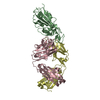
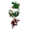
| Title | Structure of Staphylococcal Enterotoxin B bound to the neutralizing antibody 14G8 | ||||||
 Components Components |
| ||||||
 Keywords Keywords | TOXIN/IMMUNE SYSTEM / Neutralizing antibody / Staphylococcal enterotoxin B / TOXIN-IMMUNE SYSTEM complex | ||||||
| Function / homology |  Function and homology information Function and homology information | ||||||
| Biological species |   Staphylococcus aureus (bacteria) Staphylococcus aureus (bacteria) | ||||||
| Method |  X-RAY DIFFRACTION / SYNCHROTRON / MOLECULAR REPLACEMENT / Resolution: 1.8 Å X-RAY DIFFRACTION / SYNCHROTRON / MOLECULAR REPLACEMENT / Resolution: 1.8 Å | ||||||
 Authors Authors | Franklin, M.C. / Dutta, K. / Varshney, A.K. / Goger, M.J. / Fries, B.C. | ||||||
 Citation Citation | Journal: J.Biol.Chem. / Year: 2015 Title: Mechanisms mediating enhanced neutralization efficacy of staphylococcal enterotoxin B by combinations of monoclonal antibodies. Authors: Dutta, K. / Varshney, A.K. / Franklin, M.C. / Goger, M. / Wang, X. / Fries, B.C. #1: Journal: J.Biol.Chem. / Year: 2011 Title: Generation, characterization, and epitope mapping of neutralizing and protective monoclonal antibodies against staphylococcal enterotoxin B-induced lethal shock. Authors: Varshney, A.K. / Wang, X. / Cook, E. / Dutta, K. / Scharff, M.D. / Goger, M.J. / Fries, B.C. | ||||||
| History |
|
- Structure visualization
| Structure viewer | Molecule: MolmilJmol/JSmol |
|---|
- Downloads & links
-Download
| PDBx/mmCIF format | 4rgo.cif.gz | 314.2 KB | Display | PDBx/mmCIF format |
|---|---|---|---|---|
| PDB format | pdb4rgo.ent.gz | 254.7 KB | Display | PDB format |
| PDBx/mmJSON format | 4rgo.json.gz | Tree view | PDBx/mmJSON format | |
| Others |  Other downloads Other downloads |
-Validation report
| Arichive directory | https://data.pdbj.org/pub/pdb/validation_reports/rg/4rgoftp://data.pdbj.org/pub/pdb/validation_reports/rg/4rgo | HTTPS FTP |
|---|
-Related structure data
| Related structure data |  4rgmC  4rgnC 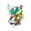 1se4S  3d9aS  3ffdS C: citing same article ( S: Starting model for refinement |
|---|---|
| Similar structure data |

-Links
 PDBj
PDBj


- Assembly
| Deposited unit | 
| ||||||||
|---|---|---|---|---|---|---|---|---|---|
| 1 |
| ||||||||
| Unit cell |
| ||||||||
| Details | The pharmacologically relevant complex contains one 14G8 Fab and one SEB monomer. There is one such complex in the asymmetric unit of the crystal. |
-Components
| #1: Protein | Mass: 28975.607 Da / Num. of mol.: 1 / Fragment: UNP residues 28-266 Source method: isolated from a genetically manipulated source Source: (gene. exp.) Staphylococcus aureus (bacteria) / Gene: entB / Production host: |
|---|---|
| #2: Antibody | Mass: 23783.713 Da / Num. of mol.: 1 / Fragment: Fab / Source method: isolated from a natural source / Source: (natural) |
| #3: Antibody | Mass: 23589.906 Da / Num. of mol.: 1 / Fragment: Fab / Source method: isolated from a natural source / Source: (natural) |
| #4: Chemical | ChemComp-ACT /   Mass: 59.044 Da / Num. of mol.: 1 / Source method: obtained synthetically / Formula: C2H3O2 Mass: 59.044 Da / Num. of mol.: 1 / Source method: obtained synthetically / Formula: C2H3O2 |
| #5: Water | ChemComp-HOH /  Mass: 18.015 Da / Num. of mol.: 908 / Source method: isolated from a natural source / Formula: H2O Mass: 18.015 Da / Num. of mol.: 908 / Source method: isolated from a natural source / Formula: H2O |
| Has protein modification | Y |
-Experimental details
-Experiment
| Experiment | Method: X-RAY DIFFRACTION / Number of used crystals: 1 |
|---|
- Sample preparation
| Crystal | Density Matthews: 2.72 Å3/Da / Density % sol: 54.8 % |
|---|---|
| Crystal grow | Temperature: 298 K / Method: vapor diffusion, sitting drop / pH: 6.5 Details: 80 mM sodium cacodylate, pH 6.5, 16% w/v PEG8000, 160 mM magnesium acetate, 20% v/v glycerol, VAPOR DIFFUSION, SITTING DROP, temperature 298K |
-Data collection
| Diffraction | Mean temperature: 100 K |
|---|---|
| Diffraction source | Source: SYNCHROTRON / Site: NSLS  / Beamline: X4C / Wavelength: 0.979 Å / Beamline: X4C / Wavelength: 0.979 Å |
| Detector | Type: MAR CCD 165 mm / Detector: CCD / Date: Feb 14, 2012 |
| Radiation | Monochromator: Si(111) / Protocol: SINGLE WAVELENGTH / Monochromatic (M) / Laue (L): M / Scattering type: x-ray |
| Radiation wavelength | Wavelength: 0.979 Å / Relative weight: 1 |
| Reflection | Resolution: 1.8→100 Å / Num. all: 77677 / Num. obs: 77133 / % possible obs: 99.3 % / Observed criterion σ(F): 0 / Observed criterion σ(I): -3 / Redundancy: 12.4 % / Rmerge(I) obs: 0.082 / Net I/σ(I): 38.5 |
| Reflection shell | Resolution: 1.8→1.83 Å / Redundancy: 4.6 % / Rmerge(I) obs: 0.561 / Mean I/σ(I) obs: 1.8 / % possible all: 89.9 |
- Processing
| Software |
| ||||||||||||||||||||||||||||||||||||||||||||||||||||||||||||||||||||||||||||||||||||||||||||||||||||||||||||||||||||||||||||||||||||||||||||||||||||||||||||||||||||||||||||||||||||||
|---|---|---|---|---|---|---|---|---|---|---|---|---|---|---|---|---|---|---|---|---|---|---|---|---|---|---|---|---|---|---|---|---|---|---|---|---|---|---|---|---|---|---|---|---|---|---|---|---|---|---|---|---|---|---|---|---|---|---|---|---|---|---|---|---|---|---|---|---|---|---|---|---|---|---|---|---|---|---|---|---|---|---|---|---|---|---|---|---|---|---|---|---|---|---|---|---|---|---|---|---|---|---|---|---|---|---|---|---|---|---|---|---|---|---|---|---|---|---|---|---|---|---|---|---|---|---|---|---|---|---|---|---|---|---|---|---|---|---|---|---|---|---|---|---|---|---|---|---|---|---|---|---|---|---|---|---|---|---|---|---|---|---|---|---|---|---|---|---|---|---|---|---|---|---|---|---|---|---|---|---|---|---|---|
| Refinement | Method to determine structure: MOLECULAR REPLACEMENT Starting model: PDB ENTRIES 1SE4, 3FFD, AND 3D9A Resolution: 1.8→42.65 Å / Cor.coef. Fo:Fc: 0.976 / Cor.coef. Fo:Fc free: 0.963 / SU B: 4.901 / SU ML: 0.068 / Cross valid method: THROUGHOUT / σ(F): 0 / ESU R: 0.154 / ESU R Free: 0.101 / Stereochemistry target values: MAXIMUM LIKELIHOOD / Details: HYDROGENS HAVE BEEN ADDED IN THE RIDING POSITIONS
| ||||||||||||||||||||||||||||||||||||||||||||||||||||||||||||||||||||||||||||||||||||||||||||||||||||||||||||||||||||||||||||||||||||||||||||||||||||||||||||||||||||||||||||||||||||||
| Solvent computation | Ion probe radii: 0.8 Å / Shrinkage radii: 0.8 Å / VDW probe radii: 1.4 Å / Solvent model: MASK | ||||||||||||||||||||||||||||||||||||||||||||||||||||||||||||||||||||||||||||||||||||||||||||||||||||||||||||||||||||||||||||||||||||||||||||||||||||||||||||||||||||||||||||||||||||||
| Displacement parameters | Biso mean: 23.132 Å2
| ||||||||||||||||||||||||||||||||||||||||||||||||||||||||||||||||||||||||||||||||||||||||||||||||||||||||||||||||||||||||||||||||||||||||||||||||||||||||||||||||||||||||||||||||||||||
| Refinement step | Cycle: LAST / Resolution: 1.8→42.65 Å
| ||||||||||||||||||||||||||||||||||||||||||||||||||||||||||||||||||||||||||||||||||||||||||||||||||||||||||||||||||||||||||||||||||||||||||||||||||||||||||||||||||||||||||||||||||||||
| Refine LS restraints |
|