 Movie
Movie Controller

[English] 日本語
 Yorodumi
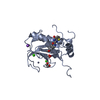
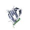
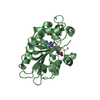
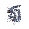
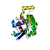
- PDB-4rcj: Crystal structure of YTHDF1 YTH domain in complex with 5mer m6A RNA -
+ Open data

- Basic information
| Entry | Database: PDB / ID: 4rcj | ||||||||||||
|---|---|---|---|---|---|---|---|---|---|---|---|---|---|
| Title | Crystal structure of YTHDF1 YTH domain in complex with 5mer m6A RNA | ||||||||||||
 Components Components |
| ||||||||||||
 Keywords Keywords | RNA BINDING PROTEIN/RNA / Structural Genomics / Structural Genomics Consortium / SGC / RNA BINDING PROTEIN-RNA complex | ||||||||||||
| Function / homology |  Function and homology information Function and homology informationregulation of axon guidance / organelle assembly / regulation of antigen processing and presentation / N6-methyladenosine-containing RNA reader activity / regulation of long-term synaptic potentiation / mRNA destabilization / positive regulation of translational initiation / immune system process / regulation of mRNA stability / stress granule assembly ...regulation of axon guidance / organelle assembly / regulation of antigen processing and presentation / N6-methyladenosine-containing RNA reader activity / regulation of long-term synaptic potentiation / mRNA destabilization / positive regulation of translational initiation / immune system process / regulation of mRNA stability / stress granule assembly / learning / positive regulation of translation / P-body / memory / cytoplasmic stress granule / ribosome binding / mRNA binding / RNA binding / cytoplasm Similarity search - Function | ||||||||||||
| Biological species |  Homo sapiens (human) Homo sapiens (human)synthetic construct (others) | ||||||||||||
| Method |  X-RAY DIFFRACTION / SYNCHROTRON / MOLECULAR REPLACEMENT / molecular replacement / Resolution: 1.6 Å X-RAY DIFFRACTION / SYNCHROTRON / MOLECULAR REPLACEMENT / molecular replacement / Resolution: 1.6 Å | ||||||||||||
 Authors Authors | Tempel, W. / Xu, C. / Bountra, C. / Arrowsmith, C.H. / Edwards, A.M. / Min, J. / Structural Genomics Consortium (SGC) | ||||||||||||
 Citation Citation | Journal: J.Biol.Chem. / Year: 2015 Title: Structural Basis for the Discriminative Recognition of N6-Methyladenosine RNA by the Human YT521-B Homology Domain Family of Proteins. Authors: Xu, C. / Liu, K. / Ahmed, H. / Loppnau, P. / Schapira, M. / Min, J. | ||||||||||||
| History |
|
- Structure visualization
| Structure viewer | Molecule: MolmilJmol/JSmol |
|---|
- Downloads & links
-Download
| PDBx/mmCIF format | 4rcj.cif.gz | 61.2 KB | Display | PDBx/mmCIF format |
|---|---|---|---|---|
| PDB format | pdb4rcj.ent.gz | 41.3 KB | Display | PDB format |
| PDBx/mmJSON format | 4rcj.json.gz | Tree view | PDBx/mmJSON format | |
| Others |  Other downloads Other downloads |
-Validation report
| Arichive directory | https://data.pdbj.org/pub/pdb/validation_reports/rc/4rcjftp://data.pdbj.org/pub/pdb/validation_reports/rc/4rcj | HTTPS FTP |
|---|
-Related structure data
| Related structure data |  4rciSC  4rcmC S: Starting model for refinement C: citing same article ( |
|---|---|
| Similar structure data |

-Links
 PDBj
PDBj- Assembly
| Deposited unit | 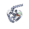
| ||||||||
|---|---|---|---|---|---|---|---|---|---|
| 1 |
| ||||||||
| Unit cell |
|
-Components
| #1: Protein | Mass: 22248.084 Da / Num. of mol.: 1 / Fragment: UNP residues 365-554 Source method: isolated from a genetically manipulated source Source: (gene. exp.) Homo sapiens (human) / Gene: YTHDF1, C20orf21 / Plasmid: pET28-MHL / Production host:  | ||||
|---|---|---|---|---|---|
| #2: RNA chain | Mass: 1600.035 Da / Num. of mol.: 1 / Source method: obtained synthetically / Source: (synth.) synthetic construct (others) | ||||
| #3: Chemical | ChemComp-UNX /   Num. of mol.: 7 / Source method: obtained synthetically Num. of mol.: 7 / Source method: obtained synthetically#4: Water | ChemComp-HOH / |  Mass: 18.015 Da / Num. of mol.: 177 / Source method: isolated from a natural source / Formula: H2O Mass: 18.015 Da / Num. of mol.: 177 / Source method: isolated from a natural source / Formula: H2OHas protein modification | N | |
-Experimental details
-Experiment
| Experiment | Method: X-RAY DIFFRACTION / Number of used crystals: 1 |
|---|
- Sample preparation
| Crystal | Density Matthews: 1.9 Å3/Da / Density % sol: 34.8 % |
|---|---|
| Crystal grow | Temperature: 293 K / Method: vapor diffusion, sitting drop / pH: 6.5 Details: 25% PEG-3350, 0.2 M sodium chloride, 0.1 M bis-tris, pH 6.5, vapor diffusion, sitting drop, temperature 293K |
-Data collection
| Diffraction | Mean temperature: 100 K | |||||||||||||||||||||
|---|---|---|---|---|---|---|---|---|---|---|---|---|---|---|---|---|---|---|---|---|---|---|
| Diffraction source | Source: SYNCHROTRON / Site: APS  / Beamline: 19-ID / Wavelength: 0.9791829 Å / Beamline: 19-ID / Wavelength: 0.9791829 Å | |||||||||||||||||||||
| Detector | Type: ADSC QUANTUM 315 / Detector: CCD / Date: Jul 17, 2014 | |||||||||||||||||||||
| Radiation | Protocol: SINGLE WAVELENGTH / Monochromatic (M) / Laue (L): M / Scattering type: x-ray | |||||||||||||||||||||
| Radiation wavelength | Wavelength: 0.9791829 Å / Relative weight: 1 | |||||||||||||||||||||
| Reflection | Resolution: 1.6→40.03 Å / Num. obs: 23138 / % possible obs: 99.4 % / Redundancy: 3.6 % / Rmerge(I) obs: 0.102 / Net I/σ(I): 9.4 | |||||||||||||||||||||
| Reflection shell | Diffraction-ID: 1
|
-Phasing
| Phasing | Method: molecular replacement |
|---|
- Processing
| Software |
| |||||||||||||||||||||||||||||||||||||||||||||||||||||||||||||||||||||||||||
|---|---|---|---|---|---|---|---|---|---|---|---|---|---|---|---|---|---|---|---|---|---|---|---|---|---|---|---|---|---|---|---|---|---|---|---|---|---|---|---|---|---|---|---|---|---|---|---|---|---|---|---|---|---|---|---|---|---|---|---|---|---|---|---|---|---|---|---|---|---|---|---|---|---|---|---|---|
| Refinement | Method to determine structure: MOLECULAR REPLACEMENT Starting model: PDB entry 4RCI Resolution: 1.6→40.03 Å / Cor.coef. Fo:Fc: 0.958 / Cor.coef. Fo:Fc free: 0.94 / WRfactor Rfree: 0.199 / WRfactor Rwork: 0.1607 / Occupancy max: 1 / Occupancy min: 0.3 / FOM work R set: 0.8468 / SU B: 2.042 / SU ML: 0.071 / SU R Cruickshank DPI: 0.0988 / SU Rfree: 0.0988 / Cross valid method: THROUGHOUT / σ(F): 0 / ESU R: 0.099 / ESU R Free: 0.099 / Stereochemistry target values: MAXIMUM LIKELIHOOD Details: ARP/WARP WAS USED IN DENSITY IMPROVEMENT MODE AFTER MOLECULAR REPLACEMENT. JLIGAND WAS USED FOR PREPARATION OF NUCLEOTIDE LINK RESTRAINT TARGETS. COOT WAS USED FOR INTERACTIVE MODEL BUILDING. ...Details: ARP/WARP WAS USED IN DENSITY IMPROVEMENT MODE AFTER MOLECULAR REPLACEMENT. JLIGAND WAS USED FOR PREPARATION OF NUCLEOTIDE LINK RESTRAINT TARGETS. COOT WAS USED FOR INTERACTIVE MODEL BUILDING. MODEL GEOMETRY WAS EVALUATED WITH MOLPROBITY. WEAK ELECTRON DENSITY NEAR THE 3'-END OF THE MODELED OLIGONUCLEOTIDE SUGGESTS THE PRESENCE OF AN ADDITIONAL NUCLEOTIDE.
| |||||||||||||||||||||||||||||||||||||||||||||||||||||||||||||||||||||||||||
| Solvent computation | Ion probe radii: 0.8 Å / Shrinkage radii: 0.8 Å / VDW probe radii: 1.2 Å / Solvent model: MASK | |||||||||||||||||||||||||||||||||||||||||||||||||||||||||||||||||||||||||||
| Displacement parameters | Biso max: 58.76 Å2 / Biso mean: 14.351 Å2 / Biso min: 4 Å2
| |||||||||||||||||||||||||||||||||||||||||||||||||||||||||||||||||||||||||||
| Refinement step | Cycle: LAST / Resolution: 1.6→40.03 Å
| |||||||||||||||||||||||||||||||||||||||||||||||||||||||||||||||||||||||||||
| Refine LS restraints |
| |||||||||||||||||||||||||||||||||||||||||||||||||||||||||||||||||||||||||||
| LS refinement shell | Resolution: 1.6→1.647 Å / Total num. of bins used: 20
|
