 Movie
Movie Controller


+ Open data

- Basic information
| Entry | Database: PDB / ID: 4rcm | |||||||||
|---|---|---|---|---|---|---|---|---|---|---|
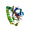
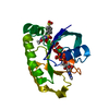
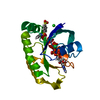
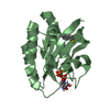
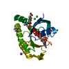
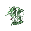
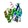
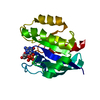
| Title | Crystal structure of the Pho92 YTH domain in complex with m6A | |||||||||
 Components Components |
| |||||||||
 Keywords Keywords | RNA BINDING PROTEIN/RNA / Structural Genomics / Structural Genomics Consortium / SGC / RNA BINDING PROTEIN-RNA complex | |||||||||
| Function / homology |  Function and homology information Function and homology information: / N6-methyladenosine-containing RNA reader activity / mRNA destabilization / regulation of mRNA stability / mRNA 3'-UTR binding / mRNA binding / cytoplasm Similarity search - Function | |||||||||
| Biological species |  synthetic construct (others) | |||||||||
| Method |  X-RAY DIFFRACTION / MOLECULAR REPLACEMENT / molecular replacement / Resolution: 1.8 Å X-RAY DIFFRACTION / MOLECULAR REPLACEMENT / molecular replacement / Resolution: 1.8 Å | |||||||||
 Authors Authors | Tempel, W. / Xu, C. / Bountra, C. / Arrowsmith, C.H. / Edwards, A.M. / Min, J. / Structural Genomics Consortium (SGC) | |||||||||
 Citation Citation | Journal: J.Biol.Chem. / Year: 2015 Title: Structural Basis for the Discriminative Recognition of N6-Methyladenosine RNA by the Human YT521-B Homology Domain Family of Proteins. Authors: Xu, C. / Liu, K. / Ahmed, H. / Loppnau, P. / Schapira, M. / Min, J. | |||||||||
| History |
|
- Structure visualization
| Structure viewer | Molecule: MolmilJmol/JSmol |
|---|
- Downloads & links
-Download
| PDBx/mmCIF format | 4rcm.cif.gz | 83.4 KB | Display | PDBx/mmCIF format |
|---|---|---|---|---|
| PDB format | pdb4rcm.ent.gz | 61.5 KB | Display | PDB format |
| PDBx/mmJSON format | 4rcm.json.gz | Tree view | PDBx/mmJSON format | |
| Others |  Other downloads Other downloads |
-Validation report
| Arichive directory | https://data.pdbj.org/pub/pdb/validation_reports/rc/4rcmftp://data.pdbj.org/pub/pdb/validation_reports/rc/4rcm | HTTPS FTP |
|---|
-Related structure data
| Related structure data |  4rciSC  4rcjC  4r3hS S: Starting model for refinement C: citing same article ( |
|---|---|
| Similar structure data |


-Links
 PDBj
PDBj- Assembly
| Deposited unit | 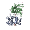
| ||||||||
|---|---|---|---|---|---|---|---|---|---|
| 1 | 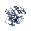
| ||||||||
| 2 | 
| ||||||||
| Unit cell |
| ||||||||
| Details | AS PER THE AUTHORS THE BIOLOGICAL ASSEMBLY IS UNKNOWN |
-Components
| #1: Protein | Mass: 19313.057 Da / Num. of mol.: 2 / Fragment: UNP residues 141-306 Source method: isolated from a genetically manipulated source Source: (gene. exp.)  #2: RNA chain | Mass: 1560.995 Da / Num. of mol.: 2 / Source method: obtained synthetically / Source: (synth.) synthetic construct (others) #3: Chemical | ChemComp-UNX /   Num. of mol.: 19 / Source method: obtained synthetically Num. of mol.: 19 / Source method: obtained synthetically#4: Water | ChemComp-HOH / |  Mass: 18.015 Da / Num. of mol.: 98 / Source method: isolated from a natural source / Formula: H2O Mass: 18.015 Da / Num. of mol.: 98 / Source method: isolated from a natural source / Formula: H2OHas protein modification | N | |
|---|
-Experimental details
-Experiment
| Experiment | Method: X-RAY DIFFRACTION / Number of used crystals: 1 |
|---|
- Sample preparation
| Crystal | Density Matthews: 1.84 Å3/Da / Density % sol: 33.29 % |
|---|---|
| Crystal grow | Temperature: 293 K / Method: vapor diffusion, sitting drop Details: 20% PEG-3350, 0.2 M potassium isocyanate, vapor diffusion, sitting drop, temperature 293K |
-Data collection
| Diffraction | Mean temperature: 100 K | ||||||||||||||||||
|---|---|---|---|---|---|---|---|---|---|---|---|---|---|---|---|---|---|---|---|
| Diffraction source | Source: ROTATING ANODE / Type: RIGAKU FR-E DW / Wavelength: 1.5418 Å | ||||||||||||||||||
| Detector | Type: RIGAKU RAXIS / Detector: IMAGE PLATE / Date: Apr 15, 2014 | ||||||||||||||||||
| Radiation | Protocol: SINGLE WAVELENGTH / Monochromatic (M) / Laue (L): M / Scattering type: x-ray | ||||||||||||||||||
| Radiation wavelength | Wavelength: 1.5418 Å / Relative weight: 1 | ||||||||||||||||||
| Reflection | Resolution: 1.8→38.4 Å / Num. obs: 27985 / % possible obs: 99.5 % / Redundancy: 3.6 % / Rmerge(I) obs: 0.09 / Net I/σ(I): 10.9 | ||||||||||||||||||
| Reflection shell | Diffraction-ID: 1 / Redundancy: 3.3 %
|
-Phasing
| Phasing | Method: molecular replacement |
|---|
- Processing
| Software |
| |||||||||||||||||||||||||||||||||||||||||||||||||||||||||||||||||||||||||||
|---|---|---|---|---|---|---|---|---|---|---|---|---|---|---|---|---|---|---|---|---|---|---|---|---|---|---|---|---|---|---|---|---|---|---|---|---|---|---|---|---|---|---|---|---|---|---|---|---|---|---|---|---|---|---|---|---|---|---|---|---|---|---|---|---|---|---|---|---|---|---|---|---|---|---|---|---|
| Refinement | Method to determine structure: MOLECULAR REPLACEMENT Starting model: PDB ENTRIES 4R3H AND 4RCI Resolution: 1.8→32.97 Å / Cor.coef. Fo:Fc: 0.937 / Cor.coef. Fo:Fc free: 0.895 / WRfactor Rfree: 0.2485 / WRfactor Rwork: 0.1999 / Occupancy max: 1 / Occupancy min: 0.3 / FOM work R set: 0.747 / SU B: 4.912 / SU ML: 0.143 / SU R Cruickshank DPI: 0.1674 / SU Rfree: 0.1578 / Cross valid method: THROUGHOUT / σ(F): 0 / ESU R: 0.167 / ESU R Free: 0.158 / Stereochemistry target values: MAXIMUM LIKELIHOOD Details: AN ENSEMBLE OF CRYSTALLOGRAPHIC MODELS OF HUMAN YTHDC1 AND YTHDF1 WAS USED AS SEARCH MODEL FOR MOLECULAR REPLACEMENT. PARROT WAS USED FOR DENSITY MODIFICATION. ARP/WARP WAS USED FOR PHASE ...Details: AN ENSEMBLE OF CRYSTALLOGRAPHIC MODELS OF HUMAN YTHDC1 AND YTHDF1 WAS USED AS SEARCH MODEL FOR MOLECULAR REPLACEMENT. PARROT WAS USED FOR DENSITY MODIFICATION. ARP/WARP WAS USED FOR PHASE IMPROVEMENT AND AUTOMATED MODEL BUILDING. COOT WAS USED FOR INTERACTIVE MODEL BUILDING. MODEL GEOMETRY WAS EVALUATED WITH MOLPROBITY.
| |||||||||||||||||||||||||||||||||||||||||||||||||||||||||||||||||||||||||||
| Solvent computation | Ion probe radii: 0.8 Å / Shrinkage radii: 0.8 Å / VDW probe radii: 1.2 Å / Solvent model: MASK | |||||||||||||||||||||||||||||||||||||||||||||||||||||||||||||||||||||||||||
| Displacement parameters | Biso max: 66.6 Å2 / Biso mean: 19.2124 Å2 / Biso min: 2 Å2
| |||||||||||||||||||||||||||||||||||||||||||||||||||||||||||||||||||||||||||
| Refinement step | Cycle: LAST / Resolution: 1.8→32.97 Å
| |||||||||||||||||||||||||||||||||||||||||||||||||||||||||||||||||||||||||||
| Refine LS restraints |
| |||||||||||||||||||||||||||||||||||||||||||||||||||||||||||||||||||||||||||
| LS refinement shell | Resolution: 1.8→1.85 Å / Total num. of bins used: 20
|
