[English] 日本語
Yorodumi
- PDB-4r8i: High Resolution Structure of a Mirror-Image RNA Oligonucleotide A... -

+
Open data


ID or keywords:

Loading...

-
Basic information

Entry
Database: PDB / ID: 4r8i
TitleHigh Resolution Structure of a Mirror-Image RNA Oligonucleotide Aptamer in Complex with the Chemokine CCL2
Components
  • C-C motif chemokine 2
  • Mirror-Image RNA Oligonucleotide Aptamer NOXE36
KeywordsCytokine/RNA / Aptamer Spiegelmer L-oligonucleotide / Cytokine-RNA complex
Function / homology
Function and homology information


helper T cell extravasation / chemokine (C-C motif) ligand 2 signaling pathway / CCR2 chemokine receptor binding / negative regulation of natural killer cell chemotaxis / astrocyte cell migration / CCR chemokine receptor binding / positive regulation of apoptotic cell clearance / negative regulation of glial cell apoptotic process / ATF4 activates genes in response to endoplasmic reticulum stress / positive regulation of glutamate receptor signaling pathway ...helper T cell extravasation / chemokine (C-C motif) ligand 2 signaling pathway / CCR2 chemokine receptor binding / negative regulation of natural killer cell chemotaxis / astrocyte cell migration / CCR chemokine receptor binding / positive regulation of apoptotic cell clearance / negative regulation of glial cell apoptotic process / ATF4 activates genes in response to endoplasmic reticulum stress / positive regulation of glutamate receptor signaling pathway / NFE2L2 regulating inflammation associated genes / chemokine-mediated signaling pathway / eosinophil chemotaxis / cellular homeostasis / negative regulation of vascular endothelial cell proliferation / chemokine activity / Chemokine receptors bind chemokines / negative regulation of G1/S transition of mitotic cell cycle / positive regulation of macrophage chemotaxis / positive regulation of calcium ion import / chemoattractant activity / macrophage chemotaxis / Interleukin-10 signaling / monocyte chemotaxis / humoral immune response / cellular response to interleukin-1 / positive regulation of endothelial cell apoptotic process / G protein-coupled receptor signaling pathway, coupled to cyclic nucleotide second messenger / cell surface receptor signaling pathway via JAK-STAT / positive regulation of synaptic transmission, glutamatergic / cellular response to fibroblast growth factor stimulus / cytoskeleton organization / sensory perception of pain / viral genome replication / animal organ morphogenesis / response to bacterium / positive regulation of T cell activation / cellular response to type II interferon / cytokine-mediated signaling pathway / chemotaxis / antimicrobial humoral immune response mediated by antimicrobial peptide / cellular response to tumor necrosis factor / regulation of cell shape / cellular response to lipopolysaccharide / angiogenesis / Interleukin-4 and Interleukin-13 signaling / negative regulation of neuron apoptotic process / cell surface receptor signaling pathway / protein phosphorylation / protein kinase activity / cell adhesion / positive regulation of cell migration / G protein-coupled receptor signaling pathway / inflammatory response / signaling receptor binding / positive regulation of gene expression / signal transduction / extracellular space / extracellular region
Similarity search - Function
CC chemokine, conserved site / Small cytokines (intercrine/chemokine) C-C subfamily signature. / Chemokine beta/gamma/delta / Intercrine alpha family (small cytokine C-X-C) (chemokine CXC). / Chemokine interleukin-8-like domain / Chemokine interleukin-8-like superfamily / Small cytokines (intecrine/chemokine), interleukin-8 like / OB fold (Dihydrolipoamide Acetyltransferase, E2P) - #40 / OB fold (Dihydrolipoamide Acetyltransferase, E2P) / Beta Barrel / Mainly Beta
Similarity search - Domain/homology
: / STRONTIUM ION / RNA / RNA (> 10) / C-C motif chemokine 2
Similarity search - Component
Biological speciesHomo sapiens (human)
synthetic construct (others)
MethodX-RAY DIFFRACTION / SYNCHROTRON / SAD / Resolution: 2.05 Å
AuthorsOberthuer, D. / Achenbach, J. / Gabdulkhakov, A. / Falke, S. / Buchner, K. / Maasch, C. / Rehders, D. / Klussmann, S. / Betzel, C.
CitationJournal: Nat Commun / Year: 2015
Title: Crystal structure of a mirror-image L-RNA aptamer (Spiegelmer) in complex with the natural L-protein target CCL2.
Authors: Oberthur, D. / Achenbach, J. / Gabdulkhakov, A. / Buchner, K. / Maasch, C. / Falke, S. / Rehders, D. / Klussmann, S. / Betzel, C.
History
DepositionSep 2, 2014Deposition site: RCSB / Processing site: RCSB
Revision 1.0Apr 29, 2015Provider: repository / Type: Initial release
Revision 1.1May 6, 2015Group: Database references
Revision 1.2Oct 30, 2024Group: Data collection / Database references ...Data collection / Database references / Derived calculations / Structure summary
Category: chem_comp_atom / chem_comp_bond ...chem_comp_atom / chem_comp_bond / database_2 / diffrn_source / pdbx_entry_details / pdbx_modification_feature / pdbx_struct_conn_angle / struct_conn / struct_ref_seq_dif / struct_site
Item: _database_2.pdbx_DOI / _database_2.pdbx_database_accession ..._database_2.pdbx_DOI / _database_2.pdbx_database_accession / _diffrn_source.pdbx_synchrotron_site / _pdbx_struct_conn_angle.ptnr1_auth_comp_id / _pdbx_struct_conn_angle.ptnr1_auth_seq_id / _pdbx_struct_conn_angle.ptnr1_label_asym_id / _pdbx_struct_conn_angle.ptnr1_label_atom_id / _pdbx_struct_conn_angle.ptnr1_label_comp_id / _pdbx_struct_conn_angle.ptnr1_label_seq_id / _pdbx_struct_conn_angle.ptnr2_auth_comp_id / _pdbx_struct_conn_angle.ptnr2_auth_seq_id / _pdbx_struct_conn_angle.ptnr2_label_asym_id / _pdbx_struct_conn_angle.ptnr2_label_atom_id / _pdbx_struct_conn_angle.ptnr2_label_comp_id / _pdbx_struct_conn_angle.ptnr3_auth_comp_id / _pdbx_struct_conn_angle.ptnr3_auth_seq_id / _pdbx_struct_conn_angle.ptnr3_label_asym_id / _pdbx_struct_conn_angle.ptnr3_label_atom_id / _pdbx_struct_conn_angle.ptnr3_label_comp_id / _pdbx_struct_conn_angle.ptnr3_label_seq_id / _pdbx_struct_conn_angle.value / _struct_conn.conn_type_id / _struct_conn.id / _struct_conn.pdbx_dist_value / _struct_conn.pdbx_leaving_atom_flag / _struct_conn.ptnr1_auth_comp_id / _struct_conn.ptnr1_auth_seq_id / _struct_conn.ptnr1_label_asym_id / _struct_conn.ptnr1_label_atom_id / _struct_conn.ptnr1_label_comp_id / _struct_conn.ptnr1_label_seq_id / _struct_conn.ptnr2_auth_comp_id / _struct_conn.ptnr2_auth_seq_id / _struct_conn.ptnr2_label_asym_id / _struct_conn.ptnr2_label_atom_id / _struct_conn.ptnr2_label_comp_id / _struct_conn.ptnr2_label_seq_id / _struct_ref_seq_dif.details / _struct_site.pdbx_auth_asym_id / _struct_site.pdbx_auth_comp_id / _struct_site.pdbx_auth_seq_id

-
Structure visualization

Structure viewerMolecule:
MolmilJmol/JSmol

Downloads & links

-
Assembly

Deposited unit
A: C-C motif chemokine 2
B: Mirror-Image RNA Oligonucleotide Aptamer NOXE36
hetero molecules


Theoretical massNumber of molelcules
Total (without water)22,0479
Polymers21,7412
Non-polymers3067
Water2,000111
1


  • Idetical with deposited unit
  • defined by author
TypeNameSymmetry operationNumber
identity operation1_555x,y,z1
Buried area1980 Å2
ΔGint-25 kcal/mol
Surface area9730 Å2
Unit cell
Length a, b, c (Å)108.912, 108.912, 34.812
Angle α, β, γ (deg.)90.00, 90.00, 90.00
Int Tables number96
Space group name H-MP43212
Components on special symmetry positions
IDModelComponents
11A-103-

HOH

Details
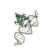
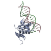
The biological assembly is a dimer generated from the complex in the asymmetric unit by the following symmetry operation: y,x,-z-2

-
Components

-
Protein / RNA chain , 2 types, 2 molecules AB

#1: Protein C-C motif chemokine 2 / HC11 / Monocyte chemoattractant protein 1 / Monocyte chemotactic and activating factor / MCAF / ...HC11 / Monocyte chemoattractant protein 1 / Monocyte chemotactic and activating factor / MCAF / Monocyte chemotactic protein 1 / MCP-1 / Monocyte secretory protein JE / Small-inducible cytokine A2


Mass: 8830.239 Da / Num. of mol.: 1
Source method: isolated from a genetically manipulated source
Source: (gene. exp.) Homo sapiens (human) / Gene: CCL2, MCP1, SCYA2 / Production host: Escherichia coli (E. coli) / References: UniProt: P13500
#2: RNA chain Mirror-Image RNA Oligonucleotide Aptamer NOXE36


Mass: 12910.646 Da / Num. of mol.: 1 / Source method: obtained synthetically
Details: The NOX-E36 L-oligonucleotides with the sequence 5'-GCACGUCCCUCACCGGUGCAAGUGAAGCCGUGGCUCUGCG-3' were synthesized using standard phosphoramidite chemistry at NOXXON Pharma AG (Berlin, Germany)
Source: (synth.) synthetic construct (others)

-
Non-polymers , 4 types, 118 molecules

#3: Chemical ChemComp-NA / SODIUM ION


Mass: 22.990 Da / Num. of mol.: 1 / Source method: obtained synthetically / Formula: Na
#4: Chemical ChemComp-SR / STRONTIUM ION


Mass: 87.620 Da / Num. of mol.: 1 / Source method: obtained synthetically / Formula: Sr
#5: Chemical
ChemComp-K / POTASSIUM ION


Mass: 39.098 Da / Num. of mol.: 5 / Source method: obtained synthetically / Formula: K
#6: Water ChemComp-HOH / water


Mass: 18.015 Da / Num. of mol.: 111 / Source method: isolated from a natural source / Formula: H2O

-
Details

Has protein modificationY

-
Experimental details

-
Experiment

ExperimentMethod: X-RAY DIFFRACTION / Number of used crystals: 1

-
Sample preparation

CrystalDensity Matthews: 2.38 Å3/Da / Density % sol: 48.38 %
Crystal growTemperature: 277 K / Method: vapor diffusion, hanging drop / pH: 5.5
Details: After thorough optimization of crystallization conditions needle shaped crystals (approx. 300x50x50um) could be obtained by mixing 1uL of the NOX-E36-CCL2 complex (in 20 mM HEPES pH 7.4, 100 ...Details: After thorough optimization of crystallization conditions needle shaped crystals (approx. 300x50x50um) could be obtained by mixing 1uL of the NOX-E36-CCL2 complex (in 20 mM HEPES pH 7.4, 100 mM NaCl, 5 mM KCl, 1 mM MgCl2, 1 mM CaCl2) with 1uL of a solution containing 40 mM Na cacodylate pH 5.5, 12 mM spermine tetrahydrochloride, 40 mM LiCl, 80 mM SrCl2 and 20 mM MgCl2 and subsequent vapor-diffusion equilibration against 1 mL of reservoir (35% (v/v) MPD, 5 mM KCl, 1 mM MgCl2 and 1 mM CaCl2). Crystals used for data collection grew within three weeks at 277K. VAPOR DIFFUSION, HANGING DROP, temperature 277K

-
Data collection

DiffractionMean temperature: 100 K
Diffraction sourceSource: SYNCHROTRON / Site: EMBL/DESY, HAMBURG / Beamline: X13 / Wavelength: 0.978 Å
DetectorType: DECTRIS PILATUS 6M / Detector: PIXEL / Date: Nov 14, 2013
RadiationMonochromator: Si / Protocol: SINGLE WAVELENGTH / Monochromatic (M) / Laue (L): M / Scattering type: x-ray
Radiation wavelengthWavelength: 0.978 Å / Relative weight: 1
ReflectionResolution: 2.05→100 Å / Num. obs: 24915 / % possible obs: 99.2 % / Observed criterion σ(F): -3 / Observed criterion σ(I): 0 / Biso Wilson estimate: 36.4 Å2

-
Processing

Software
NameVersionClassification
MxCuBEdata collection
PHENIXmodel building
REFMAC5.8.0073refinement
XDSdata reduction
XDSdata scaling
PHENIXphasing
RefinementMethod to determine structure: SAD / Resolution: 2.05→77.01 Å / Cor.coef. Fo:Fc: 0.941 / Cor.coef. Fo:Fc free: 0.925 / SU B: 4.071 / SU ML: 0.113 / Cross valid method: THROUGHOUT / ESU R: 0.215 / ESU R Free: 0.189 / Stereochemistry target values: MAXIMUM LIKELIHOOD / Details: HYDROGENS HAVE BEEN USED IF PRESENT IN THE INPUT
RfactorNum. reflection% reflectionSelection details
Rfree0.24971 1348 10.1 %RANDOM
Rwork0.20003 ---
obs0.20504 12058 97.9 %-
Solvent computationIon probe radii: 0.8 Å / Shrinkage radii: 0.8 Å / VDW probe radii: 1.2 Å / Solvent model: MASK
Displacement parametersBiso mean: 32.728 Å2
Baniso -1Baniso -2Baniso -3
1-0.21 Å2-0 Å2-0 Å2
2--0.21 Å20 Å2
3----0.41 Å2
Refinement stepCycle: LAST / Resolution: 2.05→77.01 Å
ProteinNucleic acidLigandSolventTotal
Num. atoms544 853 7 111 1515
Refine LS restraints
Refine-IDTypeDev idealDev ideal targetNumber
X-RAY DIFFRACTIONr_bond_refined_d0.0110.0231506
X-RAY DIFFRACTIONr_angle_refined_deg1.6092.6172149
X-RAY DIFFRACTIONr_dihedral_angle_1_deg5.36567
X-RAY DIFFRACTIONr_dihedral_angle_2_deg30.66224.16724
X-RAY DIFFRACTIONr_dihedral_angle_3_deg14.37815107
X-RAY DIFFRACTIONr_dihedral_angle_4_deg25.514154
X-RAY DIFFRACTIONr_chiral_restr0.0740.2279
X-RAY DIFFRACTIONr_gen_planes_refined0.0060.021813
X-RAY DIFFRACTIONr_mcbond_it2.0984.206271
X-RAY DIFFRACTIONr_mcangle_it3.6336.269337
X-RAY DIFFRACTIONr_scbond_it1.5363.0221235
X-RAY DIFFRACTIONr_long_range_B_refined5.02428.6192737
LS refinement shellResolution: 2.049→2.103 Å / Total num. of bins used: 20
RfactorNum. reflection% reflection
Rfree0.302 87 -
Rwork0.231 771 -
obs--88.91 %

+
About Yorodumi

-
News

-
Feb 9, 2022. New format data for meta-information of EMDB entries

New format data for meta-information of EMDB entries

  • Version 3 of the EMDB header file is now the official format.
  • The previous official version 1.9 will be removed from the archive.

Related info.:EMDB header

External links:wwPDB to switch to version 3 of the EMDB data model

-
Aug 12, 2020. Covid-19 info

Covid-19 info

URL: https://pdbj.org/emnavi/covid19.php

New page: Covid-19 featured information page in EM Navigator.

Related info.:Covid-19 info / Mar 5, 2020. Novel coronavirus structure data

+
Mar 5, 2020. Novel coronavirus structure data

Novel coronavirus structure data

Related info.:Yorodumi Speices / Aug 12, 2020. Covid-19 info

External links:COVID-19 featured content - PDBj / Molecule of the Month (242):Coronavirus Proteases

+
Jan 31, 2019. EMDB accession codes are about to change! (news from PDBe EMDB page)

EMDB accession codes are about to change! (news from PDBe EMDB page)

  • The allocation of 4 digits for EMDB accession codes will soon come to an end. Whilst these codes will remain in use, new EMDB accession codes will include an additional digit and will expand incrementally as the available range of codes is exhausted. The current 4-digit format prefixed with “EMD-” (i.e. EMD-XXXX) will advance to a 5-digit format (i.e. EMD-XXXXX), and so on. It is currently estimated that the 4-digit codes will be depleted around Spring 2019, at which point the 5-digit format will come into force.
  • The EM Navigator/Yorodumi systems omit the EMD- prefix.

Related info.:Q: What is EMD? / ID/Accession-code notation in Yorodumi/EM Navigator

External links:EMDB Accession Codes are Changing Soon! / Contact to PDBj

+
Jul 12, 2017. Major update of PDB

Major update of PDB

  • wwPDB released updated PDB data conforming to the new PDBx/mmCIF dictionary.
  • This is a major update changing the version number from 4 to 5, and with Remediation, in which all the entries are updated.
  • In this update, many items about electron microscopy experimental information are reorganized (e.g. em_software).
  • Now, EM Navigator and Yorodumi are based on the updated data.

External links:wwPDB Remediation / Enriched Model Files Conforming to OneDep Data Standards Now Available in the PDB FTP Archive

-
Yorodumi

Thousand views of thousand structures

  • Yorodumi is a browser for structure data from EMDB, PDB, SASBDB, etc.
  • This page is also the successor to EM Navigator detail page, and also detail information page/front-end page for Omokage search.
  • The word "yorodu" (or yorozu) is an old Japanese word meaning "ten thousand". "mi" (miru) is to see.

Related info.:EMDB / PDB / SASBDB / Comparison of 3 databanks / Yorodumi Search / Aug 31, 2016. New EM Navigator & Yorodumi / Yorodumi Papers / Jmol/JSmol / Function and homology information / Changes in new EM Navigator and Yorodumi

Read more