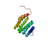
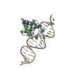
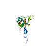
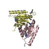
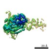
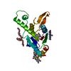
Method: simulated annealing molecluar dynamics / Software ordinal: 1 Details: THE STRUCTURES ARE BASED ON A TOTAL OF 2508 RESTRAINTS, 2263 ARE NOE-DERIVED DISTANCE RESTRAINTS 229 ARE DIHEDRAL ANGLE RESTRAINTS AND 16 ARE RESTRAINTS OF DNA BASE PLANALITY RESTRAINTS. ...Details: THE STRUCTURES ARE BASED ON A TOTAL OF 2508 RESTRAINTS, 2263 ARE NOE-DERIVED DISTANCE RESTRAINTS 229 ARE DIHEDRAL ANGLE RESTRAINTS AND 16 ARE RESTRAINTS OF DNA BASE PLANALITY RESTRAINTS. STRUCTURE CALCULATIONS WERE PERFORMED WITH CNS BY FOLLOWIN PROCEDURE. FIRST, ONLY THE STRUCTURES OF PROTEIN WERE CALCULATED USING THE HYBRID DISTANCE GEOMETRY-SIMULATED ANNEAING PROTOCOL WITH THE INTRA-PROTEIN RESTRAINTS, 10 OUT OF 100 CALCULATED STRUCTURES WERE SELECTED BY HAVING NO DISTANCE VIOLATION GREATER THAN 0.2 ANGSTROMS AND NO DIHEDRAL ANGLE VIOLATION GREATER THAN 2 DEGREES and A LOW TARGET ENERGY. SECOND, THE PROTEIN AND AN IDEALIZED B-FORM OF 16-MER DUPLEX DNA WERE DOCKED BY A SIMULATED ANNEALING PROTOCOL, FOR WHICH THE INTRA-DNA, INTER-MOLECULAR AND INTRA-PROTEIN RESTRAINTS WERE EMPLOYED. AS AN INITIAL STRUCTURE, EACH OF THE 10 PROTEIN STRUCTURES WAS PLACED IN 15 DIFFERENT POSITIONS 50 ANGSTROMS AWAY FROM THE DNA. NEXT, EACH OF THE 150 DOCKED COMPLEX STRUCTURES WAS FURTHER REFINED BY A SIMULATED ANNEALING PROTOCOL. FINALLY, 20 STRUCTURES WERE SELECTED BY HAVING NO DISTANCE VIOLATION GREATER THAN 0.2 ANGSTROME AND NO TORSION ANGLE VIOLATION THAN 2 DEGREES AND A LOW TARGET ENERGY. ADDITIONALLY, 10 CNS-SELECTED STRUCTURES WERE REFINED IN REALISTIC SOLUTION SISTEM BY USING MOLECULAR DYNAMICS SIMULATION PROGRAM, MARBLE WITH RESTRAINT FUNCTIONS FOR NOE DISTANCE DIHEDRAL ANGLE AND PLANARITY OF DNA BASE PAIR USED IN CNS.
NMR representative
Selection criteria: closest to the average
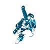
NMR ensemble
Conformer selection criteria: structures with the least restraint violations, structures with the lowest energy Conformers calculated total number: 100 / Conformers submitted total number: 10
+
About Yorodumi
-
News
-
Feb 9, 2022. New format data for meta-information of EMDB entries
New format data for meta-information of EMDB entries
Version 3 of the EMDB header file is now the official format.
The previous official version 1.9 will be removed from the archive.
In the structure databanks used in Yorodumi, some data are registered as the other names, "COVID-19 virus" and "2019-nCoV". Here are the details of the virus and the list of structure data.
Jan 31, 2019. EMDB accession codes are about to change! (news from PDBe EMDB page)
EMDB accession codes are about to change! (news from PDBe EMDB page)
The allocation of 4 digits for EMDB accession codes will soon come to an end. Whilst these codes will remain in use, new EMDB accession codes will include an additional digit and will expand incrementally as the available range of codes is exhausted. The current 4-digit format prefixed with “EMD-” (i.e. EMD-XXXX) will advance to a 5-digit format (i.e. EMD-XXXXX), and so on. It is currently estimated that the 4-digit codes will be depleted around Spring 2019, at which point the 5-digit format will come into force.
The EM Navigator/Yorodumi systems omit the EMD- prefix.
Related info.:Q: What is EMD? / ID/Accession-code notation in Yorodumi/EM Navigator
Yorodumi is a browser for structure data from EMDB, PDB, SASBDB, etc.
This page is also the successor to EM Navigator detail page, and also detail information page/front-end page for Omokage search.
The word "yorodu" (or yorozu) is an old Japanese word meaning "ten thousand". "mi" (miru) is to see.
Related info.:EMDB / PDB / SASBDB / Comparison of 3 databanks / Yorodumi Search / Aug 31, 2016. New EM Navigator & Yorodumi / Yorodumi Papers / Jmol/JSmol / Function and homology information / Changes in new EM Navigator and Yorodumi
 Movie
Movie Controller

 Yorodumi
 Open data
 Basic information
 Components
 Keywords
 Function and homology information
 Authors
 Citation
 Structure visualization
 Downloads & links
 Other downloads
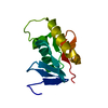


 PDBj
PDBj


 Assembly
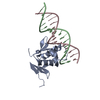
 Sample preparation
 Processing
 NMRPipe