 Movie
Movie Controller

[English] 日本語
 Yorodumi
- PDB-4r03: Crystal structure of a DUF3836 family protein (BDI_3222) from Par... -
+ Open data

- Basic information
| Entry | Database: PDB / ID: 4r03 | ||||||
|---|---|---|---|---|---|---|---|
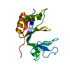
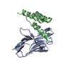
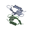
| Title | Crystal structure of a DUF3836 family protein (BDI_3222) from Parabacteroides distasonis ATCC 8503 at 1.50 A resolution | ||||||
 Components Components | Uncharacterized protein | ||||||
 Keywords Keywords | STRUCTURAL GENOMICS / UNKNOWN FUNCTION / PF12930 family protein / DUF3836 / Joint Center for Structural Genomics / JCSG / Protein Structure Initiative / PSI-BIOLOGY | ||||||
| Function / homology | Lipocalin - #720 / Protein of unknown function DUF3836 / Family of unknown function (DUF3836) / Lipocalin / Beta Barrel / Mainly Beta / CITRIC ACID / PHOSPHATE ION / DUF3836 domain-containing protein Function and homology information Function and homology information | ||||||
| Biological species |  Parabacteroides distasonis ATCC 8503 (bacteria) Parabacteroides distasonis ATCC 8503 (bacteria) | ||||||
| Method |  X-RAY DIFFRACTION / SYNCHROTRON / MAD / Resolution: 1.5 Å X-RAY DIFFRACTION / SYNCHROTRON / MAD / Resolution: 1.5 Å | ||||||
 Authors Authors | Joint Center for Structural Genomics (JCSG) | ||||||
 Citation Citation | Journal: Protein Sci. / Year: 2019 Title: Structures of single-layer beta-sheet proteins evolved from beta-hairpin repeats. Authors: Xu, Q. / Biancalana, M. / Grant, J.C. / Chiu, H.J. / Jaroszewski, L. / Knuth, M.W. / Lesley, S.A. / Godzik, A. / Elsliger, M.A. / Deacon, A.M. / Wilson, I.A. | ||||||
| History |
|
- Structure visualization
| Structure viewer | Molecule: MolmilJmol/JSmol |
|---|
- Downloads & links
-Download
| PDBx/mmCIF format | 4r03.cif.gz | 45.6 KB | Display | PDBx/mmCIF format |
|---|---|---|---|---|
| PDB format | pdb4r03.ent.gz | 31.1 KB | Display | PDB format |
| PDBx/mmJSON format | 4r03.json.gz | Tree view | PDBx/mmJSON format | |
| Others |  Other downloads Other downloads |
-Validation report
| Arichive directory | https://data.pdbj.org/pub/pdb/validation_reports/r0/4r03ftp://data.pdbj.org/pub/pdb/validation_reports/r0/4r03 | HTTPS FTP |
|---|
-Related structure data


-Links
 PDBj
PDBj- Assembly
| Deposited unit | 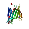
| ||||||||||||
|---|---|---|---|---|---|---|---|---|---|---|---|---|---|
| 1 |
| ||||||||||||
| Unit cell |
| ||||||||||||
| Components on special symmetry positions |
| ||||||||||||
| Details | CRYSTAL PACKING ANALYSIS SUGGESTS THE ASSIGNMENT OF A MONOMER AS THE SIGNIFICANT OLIGOMERIZATION STATE. |
-Components
| #1: Protein | Mass: 13272.422 Da / Num. of mol.: 1 / Fragment: UNP residues 29-136 Source method: isolated from a genetically manipulated source Source: (gene. exp.) Parabacteroides distasonis ATCC 8503 (bacteria)Strain: ATCC 8503 / DSM 20701 / NCTC 11152 / Gene: BDI_3222 / Plasmid: SpeedET / Production host: | ||||||
|---|---|---|---|---|---|---|---|
| #2: Chemical | ChemComp-MPD / (  Mass: 118.174 Da / Num. of mol.: 1 / Source method: obtained synthetically / Formula: C6H14O2 / Comment: precipitant*YM Mass: 118.174 Da / Num. of mol.: 1 / Source method: obtained synthetically / Formula: C6H14O2 / Comment: precipitant*YM | ||||||
| #3: Chemical | ChemComp-CIT /   Mass: 192.124 Da / Num. of mol.: 1 / Source method: obtained synthetically / Formula: C6H8O7 Mass: 192.124 Da / Num. of mol.: 1 / Source method: obtained synthetically / Formula: C6H8O7 | ||||||
| #4: Chemical |   Mass: 94.971 Da / Num. of mol.: 2 / Source method: obtained synthetically / Formula: PO4 Mass: 94.971 Da / Num. of mol.: 2 / Source method: obtained synthetically / Formula: PO4#5: Water | ChemComp-HOH / |  Mass: 18.015 Da / Num. of mol.: 188 / Source method: isolated from a natural source / Formula: H2O Mass: 18.015 Da / Num. of mol.: 188 / Source method: isolated from a natural source / Formula: H2OHas protein modification | Y | Sequence details | THIS CONSTRUCT WAS EXPRESSED WITH AN N-TERMINAL PURIFICATION TAG MGSDKIHHHHHHENLYFQG. THE TAG WAS ...THIS CONSTRUCT WAS EXPRESSED WITH AN N-TERMINAL PURIFICATI | |
-Experimental details
-Experiment
| Experiment | Method: X-RAY DIFFRACTION / Number of used crystals: 1 |
|---|
- Sample preparation
| Crystal | Density Matthews: 3.29 Å3/Da / Density % sol: 62.66 % |
|---|---|
| Crystal grow | Temperature: 277 K / Method: vapor diffusion, sitting drop / pH: 4.2 Details: 40.00% 2-methyl-2,4-pentanediol, 0.1M phosphate-citrate pH 4.2, NANODROP, VAPOR DIFFUSION, SITTING DROP, temperature 277K |
-Data collection
| Diffraction | Mean temperature: 100 K | |||||||||||||||||||||||||||||||||||||||||||||||||||||||||||||||||||||||||||||
|---|---|---|---|---|---|---|---|---|---|---|---|---|---|---|---|---|---|---|---|---|---|---|---|---|---|---|---|---|---|---|---|---|---|---|---|---|---|---|---|---|---|---|---|---|---|---|---|---|---|---|---|---|---|---|---|---|---|---|---|---|---|---|---|---|---|---|---|---|---|---|---|---|---|---|---|---|---|---|
| Diffraction source | Source: SYNCHROTRON / Site: SSRL  / Beamline: BL14-1 / Wavelength: 0.95369,0.97934,0.97907 / Beamline: BL14-1 / Wavelength: 0.95369,0.97934,0.97907 | |||||||||||||||||||||||||||||||||||||||||||||||||||||||||||||||||||||||||||||
| Detector | Type: MARMOSAIC 325 mm CCD / Detector: CCD / Date: Jun 10, 2014 Details: Vertical focusing mirror; double crystal Si(111) monochromator | |||||||||||||||||||||||||||||||||||||||||||||||||||||||||||||||||||||||||||||
| Radiation | Monochromator: double crystal Si(111) / Protocol: MAD / Monochromatic (M) / Laue (L): M / Scattering type: x-ray | |||||||||||||||||||||||||||||||||||||||||||||||||||||||||||||||||||||||||||||
| Radiation wavelength |
| |||||||||||||||||||||||||||||||||||||||||||||||||||||||||||||||||||||||||||||
| Reflection | Resolution: 1.5→27.084 Å / Num. obs: 27948 / % possible obs: 98.2 % / Observed criterion σ(I): -3 / Biso Wilson estimate: 18.114 Å2 / Rmerge(I) obs: 0.045 / Net I/σ(I): 14.96 | |||||||||||||||||||||||||||||||||||||||||||||||||||||||||||||||||||||||||||||
| Reflection shell |
|
-Phasing
| Phasing | Method: MAD |
|---|
- Processing
| Software |
| |||||||||||||||||||||||||||||||||||||||||||||||||||||||||||||||||||||||||||
|---|---|---|---|---|---|---|---|---|---|---|---|---|---|---|---|---|---|---|---|---|---|---|---|---|---|---|---|---|---|---|---|---|---|---|---|---|---|---|---|---|---|---|---|---|---|---|---|---|---|---|---|---|---|---|---|---|---|---|---|---|---|---|---|---|---|---|---|---|---|---|---|---|---|---|---|---|
| Refinement | Method to determine structure: MAD / Resolution: 1.5→27.084 Å / Cor.coef. Fo:Fc: 0.969 / Cor.coef. Fo:Fc free: 0.958 / Occupancy max: 1 / Occupancy min: 0.25 / SU B: 1.175 / SU ML: 0.042 / Cross valid method: THROUGHOUT / σ(F): 0 / ESU R: 0.061 / ESU R Free: 0.062 Stereochemistry target values: MAXIMUM LIKELIHOOD WITH PHASES Details: 1. HYDROGENS HAVE BEEN ADDED IN THE RIDING POSITIONS. 2. A MET-INHIBITION PROTOCOL WAS USED FOR SELENOMETHIONINE INCORPORATION DURING PROTEIN EXPRESSION. THE OCCUPANCY OF THE SE ATOMS IN THE ...Details: 1. HYDROGENS HAVE BEEN ADDED IN THE RIDING POSITIONS. 2. A MET-INHIBITION PROTOCOL WAS USED FOR SELENOMETHIONINE INCORPORATION DURING PROTEIN EXPRESSION. THE OCCUPANCY OF THE SE ATOMS IN THE MSE RESIDUES WAS REDUCED TO 0.75 FOR THE REDUCED SCATTERING POWER DUE TO PARTIAL S-MET INCORPORATION. 3. CITRATE (CIT), PHOSPHATE (PO4), AND 2-METHYL-2,4-PENTANEDIOL (MPD) WERE PRESENT IN PURIFICATION/CRYSTALLIZATION CONDITIONS.
| |||||||||||||||||||||||||||||||||||||||||||||||||||||||||||||||||||||||||||
| Solvent computation | Ion probe radii: 0.8 Å / Shrinkage radii: 0.8 Å / VDW probe radii: 1.2 Å / Solvent model: MASK | |||||||||||||||||||||||||||||||||||||||||||||||||||||||||||||||||||||||||||
| Displacement parameters | Biso max: 74.87 Å2 / Biso mean: 22.887 Å2 / Biso min: 9.41 Å2
| |||||||||||||||||||||||||||||||||||||||||||||||||||||||||||||||||||||||||||
| Refinement step | Cycle: LAST / Resolution: 1.5→27.084 Å
| |||||||||||||||||||||||||||||||||||||||||||||||||||||||||||||||||||||||||||
| Refine LS restraints |
| |||||||||||||||||||||||||||||||||||||||||||||||||||||||||||||||||||||||||||
| LS refinement shell | Resolution: 1.5→1.539 Å / Total num. of bins used: 20
|
