 Movie
Movie Controller


+ Open data

- Basic information
| Entry | Database: PDB / ID: 5i8j | ||||||
|---|---|---|---|---|---|---|---|
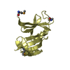
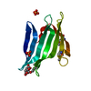
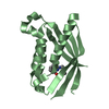
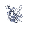
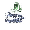
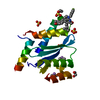
| Title | Crystal structure of Dmd from phage RB69 | ||||||
 Components Components | Dmd discriminator of mRNA degradation | ||||||
 Keywords Keywords | ANTITOXIN | ||||||
| Function / homology | Discriminator of mRNA degradation / Discriminator of mRNA degradation / Dmd discriminator of mRNA degradation Function and homology information Function and homology information | ||||||
| Biological species |  Enterobacteria phage RB69 (virus) Enterobacteria phage RB69 (virus) | ||||||
| Method |  X-RAY DIFFRACTION / SYNCHROTRON / MOLECULAR REPLACEMENT / Resolution: 1.75 Å X-RAY DIFFRACTION / SYNCHROTRON / MOLECULAR REPLACEMENT / Resolution: 1.75 Å | ||||||
 Authors Authors | Wei, Y. / Zhang, H. / Gao, Z.Q. / Dong, Y.H. | ||||||
 Citation Citation | Journal: Biochem. Biophys. Res. Commun. / Year: 2016 Title: Structural characterizations of phage antitoxin Dmd and its interactions with bacterial toxin RnlA Authors: Wei, Y. / Gao, Z.Q. / Zhang, H. / Dong, Y.H. | ||||||
| History |
|
- Structure visualization
| Structure viewer | Molecule: MolmilJmol/JSmol |
|---|
- Downloads & links
-Download
| PDBx/mmCIF format | 5i8j.cif.gz | 40.7 KB | Display | PDBx/mmCIF format |
|---|---|---|---|---|
| PDB format | pdb5i8j.ent.gz | 27.7 KB | Display | PDB format |
| PDBx/mmJSON format | 5i8j.json.gz | Tree view | PDBx/mmJSON format | |
| Others |  Other downloads Other downloads |
-Validation report
| Arichive directory | https://data.pdbj.org/pub/pdb/validation_reports/i8/5i8jftp://data.pdbj.org/pub/pdb/validation_reports/i8/5i8j | HTTPS FTP |
|---|
-Related structure data
| Related structure data |  4i8r S: Starting model for refinement |
|---|---|
| Similar structure data |


-Links
 PDBj
PDBj- Assembly
| Deposited unit | 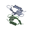
| ||||||||
|---|---|---|---|---|---|---|---|---|---|
| 1 |
| ||||||||
| Unit cell |
|
-Components
| #1: Protein | Mass: 7356.407 Da / Num. of mol.: 2 Source method: isolated from a genetically manipulated source Source: (gene. exp.) Enterobacteria phage RB69 (virus) / Gene: dmd / Production host:  #2: Water | ChemComp-HOH / |  Mass: 18.015 Da / Num. of mol.: 139 / Source method: isolated from a natural source / Formula: H2O Mass: 18.015 Da / Num. of mol.: 139 / Source method: isolated from a natural source / Formula: H2O |
|---|
-Experimental details
-Experiment
| Experiment | Method: X-RAY DIFFRACTION / Number of used crystals: 1 |
|---|
- Sample preparation
| Crystal | Density Matthews: 2.21 Å3/Da / Density % sol: 44.26 % |
|---|---|
| Crystal grow | Temperature: 293 K / Method: vapor diffusion, sitting drop / pH: 7 / Details: 0.1 M HEPES 25% PEG3350 |
-Data collection
| Diffraction | Mean temperature: 100 K |
|---|---|
| Diffraction source | Source: SYNCHROTRON / Site: BSRF  / Beamline: 1W2B / Wavelength: 0.9792 Å / Beamline: 1W2B / Wavelength: 0.9792 Å |
| Detector | Type: MAR CCD 165 mm / Detector: CCD / Date: Oct 5, 2013 |
| Radiation | Protocol: SINGLE WAVELENGTH / Monochromatic (M) / Laue (L): M / Scattering type: x-ray |
| Radiation wavelength | Wavelength: 0.9792 Å / Relative weight: 1 |
| Reflection | Resolution: 1.75→50 Å / Num. obs: 13857 / % possible obs: 99.9 % / Redundancy: 13.7 % / Rmerge(I) obs: 0.084 / Net I/σ(I): 53.76 |
| Reflection shell | Resolution: 1.75→1.78 Å / Redundancy: 11 % / Rmerge(I) obs: 0.333 / Mean I/σ(I) obs: 8.81 / % possible all: 99.3 |
- Processing
| Software |
| ||||||||||||||||||||||||||||||||||||||||||
|---|---|---|---|---|---|---|---|---|---|---|---|---|---|---|---|---|---|---|---|---|---|---|---|---|---|---|---|---|---|---|---|---|---|---|---|---|---|---|---|---|---|---|---|
| Refinement | Method to determine structure: MOLECULAR REPLACEMENT Starting model: 4I8R 4i8r Resolution: 1.75→30.914 Å / SU ML: 0.17 / Cross valid method: FREE R-VALUE / σ(F): 1.35 / Phase error: 23.36
| ||||||||||||||||||||||||||||||||||||||||||
| Solvent computation | Shrinkage radii: 0.9 Å / VDW probe radii: 1.11 Å | ||||||||||||||||||||||||||||||||||||||||||
| Refinement step | Cycle: LAST / Resolution: 1.75→30.914 Å
| ||||||||||||||||||||||||||||||||||||||||||
| Refine LS restraints |
| ||||||||||||||||||||||||||||||||||||||||||
| LS refinement shell |
|
