 Movie
Movie Controller

[English] 日本語
 Yorodumi
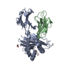
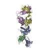
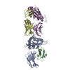
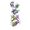
- PDB-4qrr: Crystal Structure of HLA B*3501-IPS in complex with a Delta-Beta ... -
+ Open data

- Basic information
| Entry | Database: PDB / ID: 4qrr | ||||||
|---|---|---|---|---|---|---|---|
| Title | Crystal Structure of HLA B*3501-IPS in complex with a Delta-Beta TCR, clone 12 TCR | ||||||
 Components Components |
| ||||||
 Keywords Keywords | IMMUNE SYSTEM / HLA B*3501 / human CMV / TCR / T cell | ||||||
| Function / homology |  Function and homology information Function and homology informationviral tegument / regulation of interleukin-12 production / regulation of dendritic cell differentiation / regulation of T cell anergy / regulation of interleukin-6 production / protection from natural killer cell mediated cytotoxicity / TAP binding / detection of bacterium / antigen processing and presentation of endogenous peptide antigen via MHC class Ib / antigen processing and presentation of endogenous peptide antigen via MHC class I via ER pathway, TAP-independent ...viral tegument / regulation of interleukin-12 production / regulation of dendritic cell differentiation / regulation of T cell anergy / regulation of interleukin-6 production / protection from natural killer cell mediated cytotoxicity / TAP binding / detection of bacterium / antigen processing and presentation of endogenous peptide antigen via MHC class Ib / antigen processing and presentation of endogenous peptide antigen via MHC class I via ER pathway, TAP-independent / early endosome lumen / Nef mediated downregulation of MHC class I complex cell surface expression / DAP12 interactions / secretory granule membrane / Endosomal/Vacuolar pathway / T cell mediated cytotoxicity / Antigen Presentation: Folding, assembly and peptide loading of class I MHC / lumenal side of endoplasmic reticulum membrane / regulation of iron ion transport / cellular response to iron(III) ion / negative regulation of iron ion transport / negative regulation of forebrain neuron differentiation / antigen processing and presentation of exogenous protein antigen via MHC class Ib, TAP-dependent / peptide antigen assembly with MHC class I protein complex / regulation of erythrocyte differentiation / ER to Golgi transport vesicle membrane / response to molecule of bacterial origin / defense response / HFE-transferrin receptor complex / MHC class I peptide loading complex / transferrin transport / cellular response to iron ion / negative regulation of receptor-mediated endocytosis / positive regulation of T cell cytokine production / antigen processing and presentation of endogenous peptide antigen via MHC class I / MHC class I protein complex / peptide antigen assembly with MHC class II protein complex / negative regulation of neurogenesis / cellular response to nicotine / MHC class II protein complex / positive regulation of receptor-mediated endocytosis / multicellular organismal-level iron ion homeostasis / positive regulation of T cell mediated cytotoxicity / specific granule lumen / antigen processing and presentation of exogenous peptide antigen via MHC class II / positive regulation of immune response / peptide antigen binding / phagocytic vesicle membrane / recycling endosome membrane / negative regulation of epithelial cell proliferation / positive regulation of T cell activation / Interferon gamma signaling / Immunoregulatory interactions between a Lymphoid and a non-Lymphoid cell / Interferon alpha/beta signaling / Modulation by Mtb of host immune system / sensory perception of smell / tertiary granule lumen / positive regulation of cellular senescence / MHC class II protein complex binding / T cell differentiation in thymus / DAP12 signaling / late endosome membrane / negative regulation of neuron projection development / protein refolding / protein-folding chaperone binding / ER-Phagosome pathway / early endosome membrane / structural constituent of virion / amyloid fibril formation / protein homotetramerization / host cell cytoplasm / intracellular iron ion homeostasis / adaptive immune response / learning or memory / immune response / endoplasmic reticulum lumen / Amyloid fiber formation / signaling receptor binding / Golgi membrane / external side of plasma membrane / innate immune response / lysosomal membrane / focal adhesion / Neutrophil degranulation / host cell nucleus / SARS-CoV-2 activates/modulates innate and adaptive immune responses / structural molecule activity / cell surface / Golgi apparatus / endoplasmic reticulum / protein homodimerization activity / : / extracellular exosome / extracellular region / membrane / identical protein binding / plasma membrane / cytosol Similarity search - Function | ||||||
| Biological species |  Homo sapiens (human) Homo sapiens (human)  Human herpesvirus 5 Human herpesvirus 5 | ||||||
| Method |  X-RAY DIFFRACTION / SYNCHROTRON / MOLECULAR REPLACEMENT / molecular replacement / Resolution: 3 Å X-RAY DIFFRACTION / SYNCHROTRON / MOLECULAR REPLACEMENT / molecular replacement / Resolution: 3 Å | ||||||
 Authors Authors | Gras, S. / Chabrol, E. / Rossjohn, J. | ||||||
 Citation Citation | Journal: J.Exp.Med. / Year: 2014 Title: The molecular bases of delta / alpha beta T cell-mediated antigen recognition. Authors: Pellicci, D.G. / Uldrich, A.P. / Le Nours, J. / Ross, F. / Chabrol, E. / Eckle, S.B. / de Boer, R. / Lim, R.T. / McPherson, K. / Besra, G. / Howell, A.R. / Moretta, L. / McCluskey, J. / ...Authors: Pellicci, D.G. / Uldrich, A.P. / Le Nours, J. / Ross, F. / Chabrol, E. / Eckle, S.B. / de Boer, R. / Lim, R.T. / McPherson, K. / Besra, G. / Howell, A.R. / Moretta, L. / McCluskey, J. / Heemskerk, M.H. / Gras, S. / Rossjohn, J. / Godfrey, D.I. | ||||||
| History |
|
- Structure visualization
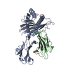
| Structure viewer | Molecule: MolmilJmol/JSmol |
|---|
- Downloads & links
-Download
| PDBx/mmCIF format | 4qrr.cif.gz | 178.1 KB | Display | PDBx/mmCIF format |
|---|---|---|---|---|
| PDB format | pdb4qrr.ent.gz | 140.3 KB | Display | PDB format |
| PDBx/mmJSON format | 4qrr.json.gz | Tree view | PDBx/mmJSON format | |
| Others |  Other downloads Other downloads |
-Validation report
| Arichive directory | https://data.pdbj.org/pub/pdb/validation_reports/qr/4qrrftp://data.pdbj.org/pub/pdb/validation_reports/qr/4qrr | HTTPS FTP |
|---|
-Related structure data
| Related structure data |  4wnqC  4wo4C  3klnS 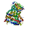 4flhS S: Starting model for refinement C: citing same article ( |
|---|---|
| Similar structure data |


-Links
 PDBj
PDBj


- Assembly
| Deposited unit | 
| ||||||||
|---|---|---|---|---|---|---|---|---|---|
| 1 |
| ||||||||
| Unit cell |
|
-Components
-Protein , 4 types, 4 molecules ABDE
| #1: Protein | Mass: 31940.246 Da / Num. of mol.: 1 Source method: isolated from a genetically manipulated source Source: (gene. exp.) Homo sapiens (human) / Gene: HLA-B, HLAB / Plasmid: pET / Production host:  |
|---|---|
| #2: Protein | Mass: 11748.160 Da / Num. of mol.: 1 Source method: isolated from a genetically manipulated source Source: (gene. exp.) Homo sapiens (human) / Gene: B2M, CDABP0092, HDCMA22P / Plasmid: pET / Production host: |
| #3: Protein | Mass: 22934.717 Da / Num. of mol.: 1 Source method: isolated from a genetically manipulated source Source: (gene. exp.) Homo sapiens (human) / Plasmid: pET / Production host: |
| #4: Protein | Mass: 27217.090 Da / Num. of mol.: 1 Source method: isolated from a genetically manipulated source Source: (gene. exp.) Homo sapiens (human) / Plasmid: pET / Production host: |
-Protein/peptide / Non-polymers , 2 types, 24 molecules P
| #5: Protein/peptide | Mass: 1081.224 Da / Num. of mol.: 1 / Source method: obtained synthetically / Source: (synth.) Human herpesvirus 5 / References: UniProt: P18139 |
|---|---|
| #6: Water | ChemComp-HOH / Mass: 18.015 Da / Num. of mol.: 23 / Source method: isolated from a natural source / Formula: H2O |
-Details
| Has protein modification | Y |
|---|
-Experimental details
-Experiment
| Experiment | Method: X-RAY DIFFRACTION / Number of used crystals: 1 |
|---|
- Sample preparation
| Crystal | Density Matthews: 2.71 Å3/Da / Density % sol: 54.68 % |
|---|---|
| Crystal grow | Temperature: 277 K / Method: vapor diffusion, hanging drop / pH: 8 Details: 0.3 M KCN, 24% PEG 3350, 2% EG, 10 mM spermidine, 10 mM L-Proline, pH 8, vapor diffusion, hanging drop, temperature 277K |
-Data collection
| Diffraction | Mean temperature: 100 K |
|---|---|
| Diffraction source | Source: SYNCHROTRON / Site: Australian Synchrotron  / Beamline: MX2 / Wavelength: 0.954 Å / Beamline: MX2 / Wavelength: 0.954 Å |
| Detector | Type: ADSC QUANTUM 315r / Detector: CCD / Date: Dec 17, 2013 |
| Radiation | Protocol: SINGLE WAVELENGTH / Monochromatic (M) / Laue (L): M / Scattering type: x-ray |
| Radiation wavelength | Wavelength: 0.954 Å / Relative weight: 1 |
| Reflection | Resolution: 3→45.95 Å / Num. all: 22376 / Num. obs: 22376 / % possible obs: 95.1 % / Redundancy: 5.1 % / Biso Wilson estimate: 62.61 Å2 / Rsym value: 0.141 |
-Phasing
| Phasing | Method: molecular replacement |
|---|
- Processing
| Software |
| ||||||||||||||||||||||||||||||||||||||||||||||||||||||||||||||||||||||||||||||||||||||||||||||||||||||||||||||||||
|---|---|---|---|---|---|---|---|---|---|---|---|---|---|---|---|---|---|---|---|---|---|---|---|---|---|---|---|---|---|---|---|---|---|---|---|---|---|---|---|---|---|---|---|---|---|---|---|---|---|---|---|---|---|---|---|---|---|---|---|---|---|---|---|---|---|---|---|---|---|---|---|---|---|---|---|---|---|---|---|---|---|---|---|---|---|---|---|---|---|---|---|---|---|---|---|---|---|---|---|---|---|---|---|---|---|---|---|---|---|---|---|---|---|---|---|
| Refinement | Method to determine structure: MOLECULAR REPLACEMENT Starting model: pdb entry 4flh, pdb entry 3kln Resolution: 3→45.95 Å / Cor.coef. Fo:Fc: 0.8697 / Cor.coef. Fo:Fc free: 0.8042 / Cross valid method: THROUGHOUT / σ(F): 0 / Stereochemistry target values: ml
| ||||||||||||||||||||||||||||||||||||||||||||||||||||||||||||||||||||||||||||||||||||||||||||||||||||||||||||||||||
| Displacement parameters | Biso mean: 51.38 Å2
| ||||||||||||||||||||||||||||||||||||||||||||||||||||||||||||||||||||||||||||||||||||||||||||||||||||||||||||||||||
| Refine analyze | Luzzati coordinate error obs: 0.445 Å | ||||||||||||||||||||||||||||||||||||||||||||||||||||||||||||||||||||||||||||||||||||||||||||||||||||||||||||||||||
| Refinement step | Cycle: LAST / Resolution: 3→45.95 Å
| ||||||||||||||||||||||||||||||||||||||||||||||||||||||||||||||||||||||||||||||||||||||||||||||||||||||||||||||||||
| Refine LS restraints |
| ||||||||||||||||||||||||||||||||||||||||||||||||||||||||||||||||||||||||||||||||||||||||||||||||||||||||||||||||||
| LS refinement shell | Resolution: 3→3.15 Å / Total num. of bins used: 11
|
