 Movie
Movie Controller

[English] 日本語
 Yorodumi
- PDB-4qih: The structure of mycobacterial glucosyl-3-phosphoglycerate phosph... -
+ Open data

- Basic information
| Entry | Database: PDB / ID: 4qih | ||||||
|---|---|---|---|---|---|---|---|
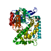
| Title | The structure of mycobacterial glucosyl-3-phosphoglycerate phosphatase Rv2419c complexes with VO3 | ||||||
 Components Components | Glucosyl-3-phosphoglycerate phosphatase | ||||||
 Keywords Keywords | HYDROLASE / glycosyl-3-phosphoglycerate / glycolysis | ||||||
| Function / homology |  Function and homology information Function and homology informationglucosyl-3-phosphoglycerate phosphatase / mannosyl-3-phosphoglycerate phosphatase / mannosyl-3-phosphoglycerate phosphatase activity Similarity search - Function | ||||||
| Biological species |   Mycobacterium tuberculosis (bacteria) Mycobacterium tuberculosis (bacteria) | ||||||
| Method |  X-RAY DIFFRACTION / SYNCHROTRON / MOLECULAR REPLACEMENT / Resolution: 2.299 Å X-RAY DIFFRACTION / SYNCHROTRON / MOLECULAR REPLACEMENT / Resolution: 2.299 Å | ||||||
 Authors Authors | Zhou, W.H. / Zheng, Q.Q. / Jiang, D.Q. / Zhang, W. / Zhang, Q.Q. / Jin, J. / Li, X. / Yang, H.T. / Shaw, N. / Rao, Z. | ||||||
 Citation Citation | Journal: J.Biol.Chem. / Year: 2014 Title: Mechanism of dephosphorylation of glucosyl-3-phosphoglycerate by a histidine phosphatase Authors: Zheng, Q. / Jiang, D. / Zhang, W. / Zhang, Q. / Zhao, Q. / Jin, J. / Li, X. / Yang, H. / Bartlam, M. / Shaw, N. / Zhou, W. / Rao, Z. | ||||||
| History |
|
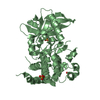
- Structure visualization
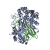
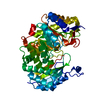
| Structure viewer | Molecule: MolmilJmol/JSmol |
|---|
- Downloads & links
-Download
| PDBx/mmCIF format | 4qih.cif.gz | 159.1 KB | Display | PDBx/mmCIF format |
|---|---|---|---|---|
| PDB format | pdb4qih.ent.gz | 127.2 KB | Display | PDB format |
| PDBx/mmJSON format | 4qih.json.gz | Tree view | PDBx/mmJSON format | |
| Others |  Other downloads Other downloads |
-Validation report
| Summary document | 4qih_validation.pdf.gz | 446.9 KB | Display | wwPDB validaton report |
|---|---|---|---|---|
| Full document | 4qih_full_validation.pdf.gz | 451 KB | Display | |
| Data in XML | 4qih_validation.xml.gz | 17.7 KB | Display | |
| Data in CIF | 4qih_validation.cif.gz | 24.6 KB | Display | |
| Arichive directory | https://data.pdbj.org/pub/pdb/validation_reports/qi/4qihftp://data.pdbj.org/pub/pdb/validation_reports/qi/4qih | HTTPS FTP |
-Related structure data
| Related structure data | 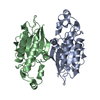 4pz9SC 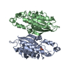 4pzaC S: Starting model for refinement C: citing same article ( |
|---|---|
| Similar structure data |


-Links
 PDBj
PDBj


- Assembly
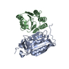
| Deposited unit | 
| ||||||||
|---|---|---|---|---|---|---|---|---|---|
| 1 |
| ||||||||
| Unit cell |
|
-Components
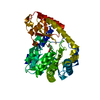
| #1: Protein | Mass: 24204.262 Da / Num. of mol.: 2 Source method: isolated from a genetically manipulated source Source: (gene. exp.) Mycobacterium tuberculosis (bacteria) / Strain: H37Rv / Gene: gpgP, MT2492, Rv2419c / Plasmid: pET28a / Production host: References: UniProt: P9WIC6, Hydrolases; Acting on ester bonds; Phosphoric-monoester hydrolases, mannosyl-3-phosphoglycerate phosphatase #2: Chemical | 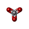  Mass: 98.940 Da / Num. of mol.: 2 / Source method: obtained synthetically / Formula: VO3 Mass: 98.940 Da / Num. of mol.: 2 / Source method: obtained synthetically / Formula: VO3#3: Water | ChemComp-HOH / |  Mass: 18.015 Da / Num. of mol.: 126 / Source method: isolated from a natural source / Formula: H2O Mass: 18.015 Da / Num. of mol.: 126 / Source method: isolated from a natural source / Formula: H2O |
|---|
-Experimental details
-Experiment
| Experiment | Method: X-RAY DIFFRACTION / Number of used crystals: 1 |
|---|
- Sample preparation
| Crystal | Density Matthews: 2.6 Å3/Da / Density % sol: 52.68 % |
|---|---|
| Crystal grow | Temperature: 293 K / Method: vapor diffusion, sitting drop / pH: 7.5 Details: 0.1M HEPES sodium pH7.5, 10% 2-propanol, 20% w/v polyethylene glycol 4000, VAPOR DIFFUSION, SITTING DROP, temperature 293K |
-Data collection
| Diffraction | Mean temperature: 100 K |
|---|---|
| Diffraction source | Source: SYNCHROTRON / Site: Photon Factory  / Beamline: BL-5A / Wavelength: 1.03935 Å / Beamline: BL-5A / Wavelength: 1.03935 Å |
| Detector | Type: ADSC QUANTUM 315r / Detector: CCD / Date: Jul 1, 2013 Details: Double crystal Monochromator with Si (111) numerical link type. Mirror 1: colli mating, Rh-coated silicon single crystal, flat shaped. Mirror 2: focusing, Rh-co ated silicon single crystal, Toroidal.roidal. |
| Radiation | Monochromator: Numerical link type Si(111) double crystal monochromator, direct water cooling using micro-channel (1st crystal), indirect water cooling (2nd crystal) Protocol: SINGLE WAVELENGTH / Monochromatic (M) / Laue (L): M / Scattering type: x-ray |
| Radiation wavelength | Wavelength: 1.03935 Å / Relative weight: 1 |
| Reflection | Resolution: 2.299→50 Å / Num. all: 23222 / Num. obs: 22409 / % possible obs: 96.5 % / Observed criterion σ(F): 1 / Observed criterion σ(I): 1 / Redundancy: 7 % / Rmerge(I) obs: 0.055 / Rsym value: 0.05 / Net I/σ(I): 46 |
| Reflection shell | Resolution: 2.299→2.34 Å / Redundancy: 7 % / Rmerge(I) obs: 0.27 / Mean I/σ(I) obs: 9 / Num. unique all: 1128 / Rsym value: 0.22 / % possible all: 100 |
- Processing
| Software |
| ||||||||||||||||||||||||||||||||||||||||||||||||||||||||||||||||||||||||||||||||||||||||||
|---|---|---|---|---|---|---|---|---|---|---|---|---|---|---|---|---|---|---|---|---|---|---|---|---|---|---|---|---|---|---|---|---|---|---|---|---|---|---|---|---|---|---|---|---|---|---|---|---|---|---|---|---|---|---|---|---|---|---|---|---|---|---|---|---|---|---|---|---|---|---|---|---|---|---|---|---|---|---|---|---|---|---|---|---|---|---|---|---|---|---|---|
| Refinement | Method to determine structure: MOLECULAR REPLACEMENT Starting model: 4PZ9 Resolution: 2.299→35.008 Å / SU ML: 0.26 / Cross valid method: THROUGHOUT / σ(F): 1.34 / σ(I): 0 / Phase error: 25.43 / Stereochemistry target values: ML
| ||||||||||||||||||||||||||||||||||||||||||||||||||||||||||||||||||||||||||||||||||||||||||
| Solvent computation | Shrinkage radii: 0.9 Å / VDW probe radii: 1.11 Å / Solvent model: FLAT BULK SOLVENT MODEL | ||||||||||||||||||||||||||||||||||||||||||||||||||||||||||||||||||||||||||||||||||||||||||
| Refinement step | Cycle: LAST / Resolution: 2.299→35.008 Å
| ||||||||||||||||||||||||||||||||||||||||||||||||||||||||||||||||||||||||||||||||||||||||||
| Refine LS restraints |
| ||||||||||||||||||||||||||||||||||||||||||||||||||||||||||||||||||||||||||||||||||||||||||
| LS refinement shell | Refine-ID: X-RAY DIFFRACTION / Total num. of bins used: 14
|
