[English] 日本語
 Yorodumi
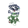
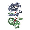
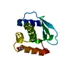
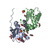
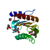
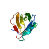
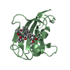
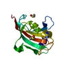
- PDB-4qhf: Crystal structure of Methanocaldococcus jannaschii monomeric selecase -
+ Open data
- Basic information
| Entry | Database: PDB / ID: 4qhf | ||||||
|---|---|---|---|---|---|---|---|
| Title | Crystal structure of Methanocaldococcus jannaschii monomeric selecase | ||||||
 Components Components | Uncharacterized protein MJ1213 | ||||||
 Keywords Keywords | HYDROLASE / Minigluzincin / Proteolytic enzyme | ||||||
| Function / homology | Metalloproteases ("zincins"), catalytic domain / Peptidase M56 / BlaR1 peptidase M56 / Zincin-like / 2-Layer Sandwich / Alpha Beta / NICKEL (II) ION / Uncharacterized protein MJ1213 Function and homology information Function and homology information | ||||||
| Biological species |   Methanocaldococcus jannaschii (archaea) Methanocaldococcus jannaschii (archaea) | ||||||
| Method |  X-RAY DIFFRACTION / X-RAY DIFFRACTION /  SYNCHROTRON / SYNCHROTRON /  MOLECULAR REPLACEMENT / Resolution: 2.1 Å MOLECULAR REPLACEMENT / Resolution: 2.1 Å | ||||||
 Authors Authors | Lopez-pelegrin, M. / Cerda-costa, N. / Cintas-pedrola, A. / Herranz-trillo, F. / Bernado, P. / Peinado, J.R. / Arolas, J.L. / Gomis-ruth, F.X. | ||||||
 Citation Citation |  Journal: Angew.Chem.Int.Ed.Engl. / Year: 2014 Journal: Angew.Chem.Int.Ed.Engl. / Year: 2014Title: Multiple stable conformations account for reversible concentration-dependent oligomerization and autoinhibition of a metamorphic metallopeptidase Authors: Lopez-Pelegrin, M. / Cerda-Costa, N. / Cintas-Pedrola, A. / Herranz-Trillo, F. / Bernado, P. / Peinado, J.R. / Arolas, J.L. / Gomis-Ruth, F.X. | ||||||
| History |
|
- Structure visualization
| Structure viewer | Molecule:  Molmil Molmil Jmol/JSmol Jmol/JSmol |
|---|
- Downloads & links
- Download
| PDBx/mmCIF format |  4qhf.cif.gz 4qhf.cif.gz | 60.5 KB | Display |  PDBx/mmCIF format PDBx/mmCIF format |
|---|---|---|---|---|
| PDB format |  pdb4qhf.ent.gz pdb4qhf.ent.gz | 44.9 KB | Display |  PDB format PDB format |
| PDBx/mmJSON format |  4qhf.json.gz 4qhf.json.gz | Tree view |  PDBx/mmJSON format PDBx/mmJSON format | |
| Others |  Other downloads Other downloads |
-Validation report
| Summary document |  4qhf_validation.pdf.gz 4qhf_validation.pdf.gz | 438.8 KB | Display |  wwPDB validaton report wwPDB validaton report |
|---|---|---|---|---|
| Full document |  4qhf_full_validation.pdf.gz 4qhf_full_validation.pdf.gz | 438.8 KB | Display | |
| Data in XML |  4qhf_validation.xml.gz 4qhf_validation.xml.gz | 7 KB | Display | |
| Data in CIF |  4qhf_validation.cif.gz 4qhf_validation.cif.gz | 8.7 KB | Display | |
| Arichive directory |  https://data.pdbj.org/pub/pdb/validation_reports/qh/4qhf https://data.pdbj.org/pub/pdb/validation_reports/qh/4qhf ftp://data.pdbj.org/pub/pdb/validation_reports/qh/4qhf ftp://data.pdbj.org/pub/pdb/validation_reports/qh/4qhf | HTTPS FTP |
-Related structure data
- Links
Links
- Assembly
| Deposited unit | 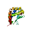
| |||||||||
|---|---|---|---|---|---|---|---|---|---|---|
| 1 |
| |||||||||
| Unit cell |
| |||||||||
| Components on special symmetry positions |
|
- Components
| #1: Protein | Mass: 13103.546 Da / Num. of mol.: 1 Source method: isolated from a genetically manipulated source Source: (gene. exp.)   Methanocaldococcus jannaschii (archaea) Methanocaldococcus jannaschii (archaea)Strain: ATCC 43067 / DSM 2661 / JAL-1 / JCM 10045 / NBRC 100440 Gene: MJ1213 / Plasmid: pET28 / Production host:  |
|---|---|
| #2: Chemical | ChemComp-NI / |
| #3: Chemical | ChemComp-GOL / |
| #4: Water | ChemComp-HOH / |
-Experimental details
-Experiment
| Experiment | Method:  X-RAY DIFFRACTION / Number of used crystals: 1 X-RAY DIFFRACTION / Number of used crystals: 1 |
|---|
- Sample preparation
| Crystal | Density Matthews: 2.35 Å3/Da / Density % sol: 47.73 % |
|---|---|
| Crystal grow | Temperature: 293 K / Method: vapor diffusion, sitting drop / pH: 8.5 Details: 0.1M Tris-HCl, 0.2M sodium acetate, 30%(w/v) polyethylene glycol 4,000, pH 8.5, VAPOR DIFFUSION, SITTING DROP, temperature 293K |
-Data collection
| Diffraction | Mean temperature: 100 K |
|---|---|
| Diffraction source | Source:  SYNCHROTRON / Site: SYNCHROTRON / Site:  ESRF ESRF  / Beamline: ID23-2 / Wavelength: 0.8726 Å / Beamline: ID23-2 / Wavelength: 0.8726 Å |
| Detector | Type: ADSC QUANTUM 315r / Detector: CCD / Date: Jun 19, 2013 |
| Radiation | Monochromator: Si(311) / Protocol: SINGLE WAVELENGTH / Monochromatic (M) / Laue (L): M / Scattering type: x-ray |
| Radiation wavelength | Wavelength: 0.8726 Å / Relative weight: 1 |
| Reflection | Resolution: 2.1→42.7 Å / Num. all: 7658 / Num. obs: 7658 / % possible obs: 99.9 % / Observed criterion σ(F): 0 / Observed criterion σ(I): 0 / Biso Wilson estimate: 37.95 Å2 / Rmerge(I) obs: 0.07 / Net I/σ(I): 17.7 |
| Reflection shell | Resolution: 2.1→42.7 Å / Rmerge(I) obs: 0.07 / Mean I/σ(I) obs: 17.7 / Num. unique all: 7658 / % possible all: 99.9 |
- Processing
| Software |
| |||||||||||||||||||||||||||||||||||||||||||||||||||||||||||||||||||||||||||
|---|---|---|---|---|---|---|---|---|---|---|---|---|---|---|---|---|---|---|---|---|---|---|---|---|---|---|---|---|---|---|---|---|---|---|---|---|---|---|---|---|---|---|---|---|---|---|---|---|---|---|---|---|---|---|---|---|---|---|---|---|---|---|---|---|---|---|---|---|---|---|---|---|---|---|---|---|
| Refinement | Method to determine structure:  MOLECULAR REPLACEMENT MOLECULAR REPLACEMENTStarting model: Dimeric selecase Resolution: 2.1→42.7 Å / Cor.coef. Fo:Fc: 0.9401 / Cor.coef. Fo:Fc free: 0.9134 / SU R Cruickshank DPI: 0.223 / Cross valid method: THROUGHOUT / σ(F): 0 / Stereochemistry target values: Engh & Huber
| |||||||||||||||||||||||||||||||||||||||||||||||||||||||||||||||||||||||||||
| Displacement parameters | Biso mean: 50.25 Å2
| |||||||||||||||||||||||||||||||||||||||||||||||||||||||||||||||||||||||||||
| Refine analyze | Luzzati coordinate error obs: 0.304 Å | |||||||||||||||||||||||||||||||||||||||||||||||||||||||||||||||||||||||||||
| Refinement step | Cycle: LAST / Resolution: 2.1→42.7 Å
| |||||||||||||||||||||||||||||||||||||||||||||||||||||||||||||||||||||||||||
| Refine LS restraints |
| |||||||||||||||||||||||||||||||||||||||||||||||||||||||||||||||||||||||||||
| LS refinement shell | Resolution: 2.1→2.2 Å / Total num. of bins used: 5
| |||||||||||||||||||||||||||||||||||||||||||||||||||||||||||||||||||||||||||
| Refinement TLS params. | Method: refined / Refine-ID: X-RAY DIFFRACTION
| |||||||||||||||||||||||||||||||||||||||||||||||||||||||||||||||||||||||||||
| Refinement TLS group |
|
 Movie
Movie Controller




 PDBj
PDBj
