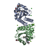
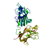
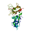
[English] 日本語
Yorodumi
- PDB-4qdr: Physical basis for Nrp2 ligand binding -

+
Open data


ID or keywords:

Loading...

-
Basic information

Entry
Database: PDB / ID: 4qdr
TitlePhysical basis for Nrp2 ligand binding
ComponentsNeuropilin-2Neuropilin
KeywordsCELL ADHESION / coagulation factor domain / discoidin domain / receptor / VEGF-C / membrane
Function / homology
Function and homology information


vestibulocochlear nerve structural organization / dorsal root ganglion morphogenesis / ventral trunk neural crest cell migration / sympathetic neuron projection guidance / facioacoustic ganglion development / trigeminal ganglion development / trigeminal nerve structural organization / sensory neuron axon guidance / facial nerve structural organization / gonadotrophin-releasing hormone neuronal migration to the hypothalamus ...vestibulocochlear nerve structural organization / dorsal root ganglion morphogenesis / ventral trunk neural crest cell migration / sympathetic neuron projection guidance / facioacoustic ganglion development / trigeminal ganglion development / trigeminal nerve structural organization / sensory neuron axon guidance / facial nerve structural organization / gonadotrophin-releasing hormone neuronal migration to the hypothalamus / branchiomotor neuron axon guidance / axon extension involved in axon guidance / NrCAM interactions / sympathetic neuron projection extension / Neurophilin interactions with VEGF and VEGFR / sympathetic ganglion development / neural crest cell migration involved in autonomic nervous system development / vascular endothelial growth factor receptor activity / nerve development / semaphorin receptor complex / outflow tract septum morphogenesis / semaphorin receptor activity / regulation of postsynapse organization / negative chemotaxis / cytokine binding / growth factor binding / semaphorin-plexin signaling pathway / positive regulation of endothelial cell proliferation / cellular response to leukemia inhibitory factor / positive regulation of endothelial cell migration / axon guidance / signaling receptor activity / heparin binding / postsynaptic membrane / angiogenesis / cell adhesion / axon / glutamatergic synapse / extracellular region / membrane / identical protein binding / metal ion binding / plasma membrane
Similarity search - Function
Neuropilin / Neuropilin, C-terminal / C-terminal domain of neuropilin glycoprotein / Domain in meprin, A5, receptor protein tyrosine phosphatase mu (and others) / MAM domain, meprin/A5/mu / MAM domain / Coagulation factors 5/8 type C domain (FA58C) signature 2. / MAM domain profile. / CUB domain / Domain first found in C1r, C1s, uEGF, and bone morphogenetic protein. ...Neuropilin / Neuropilin, C-terminal / C-terminal domain of neuropilin glycoprotein / Domain in meprin, A5, receptor protein tyrosine phosphatase mu (and others) / MAM domain, meprin/A5/mu / MAM domain / Coagulation factors 5/8 type C domain (FA58C) signature 2. / MAM domain profile. / CUB domain / Domain first found in C1r, C1s, uEGF, and bone morphogenetic protein. / CUB domain / CUB domain profile. / Coagulation factors 5/8 type C domain (FA58C) signature 1. / Spermadhesin, CUB domain superfamily / Coagulation factor 5/8 C-terminal domain, discoidin domain / Coagulation factors 5/8 type C domain (FA58C) profile. / F5/8 type C domain / Coagulation factor 5/8 C-terminal domain / Galactose-binding domain-like / Galactose-binding-like domain superfamily / Concanavalin A-like lectin/glucanase domain superfamily / Jelly Rolls / Sandwich / Mainly Beta
Similarity search - Domain/homology
Biological speciesHomo sapiens (human)
MethodX-RAY DIFFRACTION / SYNCHROTRON / MOLECULAR REPLACEMENT / Resolution: 2.4 Å
AuthorsParker, M.W. / Vander Kooi, C.W.
CitationJournal: Structure / Year: 2015
Title: Structural Basis for VEGF-C Binding to Neuropilin-2 and Sequestration by a Soluble Splice Form.
Authors: Parker, M.W. / Linkugel, A.D. / Goel, H.L. / Wu, T. / Mercurio, A.M. / Vander Kooi, C.W.
History
DepositionMay 14, 2014Deposition site: RCSB / Processing site: RCSB
Revision 1.0Apr 15, 2015Provider: repository / Type: Initial release
Revision 1.1May 6, 2015Group: Database references
Revision 1.2Sep 20, 2023Group: Data collection / Database references / Refinement description
Category: chem_comp_atom / chem_comp_bond ...chem_comp_atom / chem_comp_bond / database_2 / pdbx_initial_refinement_model / struct_ref_seq_dif
Item: _database_2.pdbx_DOI / _database_2.pdbx_database_accession / _struct_ref_seq_dif.details

-
Structure visualization

Structure viewerMolecule:
MolmilJmol/JSmol

Downloads & links

-
Assembly

Deposited unit
A: Neuropilin-2


Theoretical massNumber of molelcules
Total (without water)36,5991
Polymers36,5991
Non-polymers00
Water2,216123
1


  • Idetical with deposited unit
  • defined by author&software
TypeNameSymmetry operationNumber
identity operation1_555x,y,z1
MethodPISA
Unit cell
Length a, b, c (Å)34.901, 70.759, 122.974
Angle α, β, γ (deg.)90.00, 90.00, 90.00
Int Tables number19
Space group name H-MP212121

-
Components

#1: Protein Neuropilin-2 / Neuropilin / Vascular endothelial cell growth factor 165 receptor 2


Mass: 36599.215 Da / Num. of mol.: 1 / Fragment: unp residues 276-595 / Mutation: T319R
Source method: isolated from a genetically manipulated source
Source: (gene. exp.) Homo sapiens (human) / Gene: NRP2, VEGF165R2 / Plasmid: pET28b / Production host: Escherichia coli (E. coli) / Strain (production host): Rosetta-gami 2(DE3) / References: UniProt: O60462
#2: Water ChemComp-HOH / water / Water


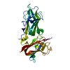
Mass: 18.015 Da / Num. of mol.: 123 / Source method: isolated from a natural source / Formula: H2O

-
Experimental details

-
Experiment

ExperimentMethod: X-RAY DIFFRACTION / Number of used crystals: 1

-
Sample preparation

CrystalDensity Matthews: 2.07 Å3/Da / Density % sol: 40.71 %
Crystal growTemperature: 298 K / Method: vapor diffusion, hanging drop / pH: 7
Details: 0.1 M HEPES, 18% (w/v) PEG 12000, pH 7, VAPOR DIFFUSION, HANGING DROP, temperature 298K

-
Data collection

DiffractionMean temperature: 110 K
Diffraction sourceSource: SYNCHROTRON / Site: APS / Beamline: 22-BM / Wavelength: 1 Å
DetectorType: MARMOSAIC 225 mm CCD / Detector: CCD / Date: Jun 25, 2011
RadiationMonochromator: Si(111) / Protocol: SINGLE WAVELENGTH / Monochromatic (M) / Laue (L): M / Scattering type: x-ray
Radiation wavelengthWavelength: 1 Å / Relative weight: 1
ReflectionResolution: 2.4→20 Å / Num. all: 12715 / Num. obs: 12223 / % possible obs: 96.4 % / Observed criterion σ(I): -3 / Redundancy: 29.4 % / Rmerge(I) obs: 0.08
Reflection shellResolution: 2.4→2.49 Å / Redundancy: 5.1 % / Rmerge(I) obs: 0.292 / % possible all: 83.2

-
Processing

Software
NameVersionClassification
HKL-2000data collection
MOLREPphasing
REFMAC5.6.0117refinement
HKL-2000data reduction
HKL-2000data scaling
RefinementMethod to determine structure: MOLECULAR REPLACEMENT
Starting model: pdb entry 2QQJ
Resolution: 2.4→19.69 Å / Cor.coef. Fo:Fc: 0.948 / Cor.coef. Fo:Fc free: 0.926 / SU B: 18.199 / SU ML: 0.231 / Cross valid method: THROUGHOUT / ESU R: 0.829 / ESU R Free: 0.302 / Stereochemistry target values: MAXIMUM LIKELIHOOD / Details: HYDROGENS HAVE BEEN USED IF PRESENT IN THE INPUT
RfactorNum. reflection% reflectionSelection details
Rfree0.25512 586 4.9 %RANDOM
Rwork0.20089 ---
obs0.20357 11490 96.26 %-
Solvent computationIon probe radii: 0.8 Å / Shrinkage radii: 0.8 Å / VDW probe radii: 1.2 Å / Solvent model: MASK
Displacement parametersBiso mean: 46.599 Å2
Baniso -1Baniso -2Baniso -3
1--0.34 Å20 Å20 Å2
2--0.09 Å2-0 Å2
3---0.25 Å2
Refinement stepCycle: LAST / Resolution: 2.4→19.69 Å
ProteinNucleic acidLigandSolventTotal
Num. atoms2513 0 0 123 2636
Refine LS restraints
Refine-IDTypeDev idealDev ideal targetNumber
X-RAY DIFFRACTIONr_bond_refined_d0.0080.0192579
X-RAY DIFFRACTIONr_bond_other_d
X-RAY DIFFRACTIONr_angle_refined_deg1.191.9383500
X-RAY DIFFRACTIONr_angle_other_deg
X-RAY DIFFRACTIONr_dihedral_angle_1_deg6.9965312
X-RAY DIFFRACTIONr_dihedral_angle_2_deg37.18323.228127
X-RAY DIFFRACTIONr_dihedral_angle_3_deg14.16315428
X-RAY DIFFRACTIONr_dihedral_angle_4_deg17.9421524
X-RAY DIFFRACTIONr_chiral_restr0.080.2366
X-RAY DIFFRACTIONr_gen_planes_refined0.0040.0212001
X-RAY DIFFRACTIONr_gen_planes_other
X-RAY DIFFRACTIONr_nbd_refined
X-RAY DIFFRACTIONr_nbd_other
X-RAY DIFFRACTIONr_nbtor_refined
X-RAY DIFFRACTIONr_nbtor_other
X-RAY DIFFRACTIONr_xyhbond_nbd_refined
X-RAY DIFFRACTIONr_xyhbond_nbd_other
X-RAY DIFFRACTIONr_metal_ion_refined
X-RAY DIFFRACTIONr_metal_ion_other
X-RAY DIFFRACTIONr_symmetry_vdw_refined
X-RAY DIFFRACTIONr_symmetry_vdw_other
X-RAY DIFFRACTIONr_symmetry_hbond_refined
X-RAY DIFFRACTIONr_symmetry_hbond_other
X-RAY DIFFRACTIONr_symmetry_metal_ion_refined
X-RAY DIFFRACTIONr_symmetry_metal_ion_other
X-RAY DIFFRACTIONr_mcbond_it
X-RAY DIFFRACTIONr_mcbond_other
X-RAY DIFFRACTIONr_mcangle_it
X-RAY DIFFRACTIONr_mcangle_other
X-RAY DIFFRACTIONr_scbond_it
X-RAY DIFFRACTIONr_scbond_other
X-RAY DIFFRACTIONr_scangle_it
X-RAY DIFFRACTIONr_scangle_other
X-RAY DIFFRACTIONr_long_range_B_refined
X-RAY DIFFRACTIONr_long_range_B_other
X-RAY DIFFRACTIONr_rigid_bond_restr
X-RAY DIFFRACTIONr_sphericity_free
X-RAY DIFFRACTIONr_sphericity_bonded
LS refinement shellResolution: 2.4→2.461 Å / Total num. of bins used: 20
RfactorNum. reflection% reflection
Rfree0.487 26 -
Rwork0.286 633 -
obs--81.36 %
Refinement TLS params.Method: refined / Origin x: 1.752 Å / Origin y: 7.713 Å / Origin z: -14.935 Å
111213212223313233
T0.321 Å2-0.0648 Å20.0041 Å2-0.2817 Å2-0.0157 Å2--0.1616 Å2
L0.2769 °20.2614 °2-0.0364 °2-3.1313 °2-0.9582 °2--1.3307 °2
S0.0504 Å °-0.0062 Å °0.0149 Å °0.2341 Å °-0.0663 Å °-0.0474 Å °-0.2988 Å °0.1781 Å °0.0159 Å °
Refinement TLS group
IDRefine-IDRefine TLS-IDAuth asym-IDAuth seq-ID
1X-RAY DIFFRACTION1A274 - 594
2X-RAY DIFFRACTION1A601 - 723

+
About Yorodumi

-
News

-
Feb 9, 2022. New format data for meta-information of EMDB entries

New format data for meta-information of EMDB entries

  • Version 3 of the EMDB header file is now the official format.
  • The previous official version 1.9 will be removed from the archive.

Related info.:EMDB header

External links:wwPDB to switch to version 3 of the EMDB data model

-
Aug 12, 2020. Covid-19 info

Covid-19 info

URL: https://pdbj.org/emnavi/covid19.php

New page: Covid-19 featured information page in EM Navigator.

Related info.:Covid-19 info / Mar 5, 2020. Novel coronavirus structure data

+
Mar 5, 2020. Novel coronavirus structure data

Novel coronavirus structure data

Related info.:Yorodumi Speices / Aug 12, 2020. Covid-19 info

External links:COVID-19 featured content - PDBj / Molecule of the Month (242):Coronavirus Proteases

+
Jan 31, 2019. EMDB accession codes are about to change! (news from PDBe EMDB page)

EMDB accession codes are about to change! (news from PDBe EMDB page)

  • The allocation of 4 digits for EMDB accession codes will soon come to an end. Whilst these codes will remain in use, new EMDB accession codes will include an additional digit and will expand incrementally as the available range of codes is exhausted. The current 4-digit format prefixed with “EMD-” (i.e. EMD-XXXX) will advance to a 5-digit format (i.e. EMD-XXXXX), and so on. It is currently estimated that the 4-digit codes will be depleted around Spring 2019, at which point the 5-digit format will come into force.
  • The EM Navigator/Yorodumi systems omit the EMD- prefix.

Related info.:Q: What is EMD? / ID/Accession-code notation in Yorodumi/EM Navigator

External links:EMDB Accession Codes are Changing Soon! / Contact to PDBj

+
Jul 12, 2017. Major update of PDB

Major update of PDB

  • wwPDB released updated PDB data conforming to the new PDBx/mmCIF dictionary.
  • This is a major update changing the version number from 4 to 5, and with Remediation, in which all the entries are updated.
  • In this update, many items about electron microscopy experimental information are reorganized (e.g. em_software).
  • Now, EM Navigator and Yorodumi are based on the updated data.

External links:wwPDB Remediation / Enriched Model Files Conforming to OneDep Data Standards Now Available in the PDB FTP Archive

-
Yorodumi

Thousand views of thousand structures

  • Yorodumi is a browser for structure data from EMDB, PDB, SASBDB, etc.
  • This page is also the successor to EM Navigator detail page, and also detail information page/front-end page for Omokage search.
  • The word "yorodu" (or yorozu) is an old Japanese word meaning "ten thousand". "mi" (miru) is to see.

Related info.:EMDB / PDB / SASBDB / Comparison of 3 databanks / Yorodumi Search / Aug 31, 2016. New EM Navigator & Yorodumi / Yorodumi Papers / Jmol/JSmol / Function and homology information / Changes in new EM Navigator and Yorodumi

Read more