| Entry | Database: PDB / ID: 4qb5
|
|---|
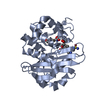
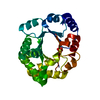
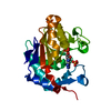
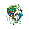
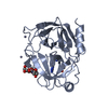
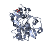
| Title | Crystal structure of a glyoxalase/bleomycin resistance protein from Albidiferax ferrireducens T118 |
|---|
 Components Components | Glyoxalase/bleomycin resistance protein/dioxygenase |
|---|
 Keywords Keywords | OXIDOREDUCTASE / Structural Genomics / PSI-Biology / New York Structural Genomics Research Consortium / NYSGRC / PROTEIN STRUCTURE INITIATIVE / alpha/beta |
|---|
| Function / homology |  Function and homology information Function and homology information
2,3-Dihydroxybiphenyl 1,2-Dioxygenase, domain 1 / 2,3-Dihydroxybiphenyl 1,2-Dioxygenase; domain 1 / Glyoxalase/fosfomycin resistance/dioxygenase domain / Glyoxalase/Bleomycin resistance protein/Dioxygenase superfamily / Vicinal oxygen chelate (VOC) domain / Vicinal oxygen chelate (VOC) domain profile. / Glyoxalase/Bleomycin resistance protein/Dihydroxybiphenyl dioxygenase / Roll / Alpha BetaSimilarity search - Domain/homology |
|---|
| Biological species |  Albidiferax ferrireducens T118 (bacteria) Albidiferax ferrireducens T118 (bacteria) |
|---|
| Method |  X-RAY DIFFRACTION / SYNCHROTRON / SAD / Resolution: 2.05 Å X-RAY DIFFRACTION / SYNCHROTRON / SAD / Resolution: 2.05 Å |
|---|
 Authors Authors | Kumaran, D. / Chamala, S. / Evans, B. / Foti, R. / Gizzi, A. / Hillerich, B. / Kar, A. / Lafleur, J. / Seidel, R. / Villigas, G. ...Kumaran, D. / Chamala, S. / Evans, B. / Foti, R. / Gizzi, A. / Hillerich, B. / Kar, A. / Lafleur, J. / Seidel, R. / Villigas, G. / Zencheck, W. / Al Obaidi, N. / Almo, S.C. / Swaminathan, S. / New York Structural Genomics Research Consortium (NYSGRC) |
|---|
 Citation Citation | Journal: To be Published
Title: Crystal structure of a glyoxalase/bleomycin resistance protein from Albidiferax ferrireducens T118
Authors: Kumaran, D. / Almo, S.C. / Swaminathan, S. |
|---|
| History | | Deposition | May 6, 2014 | Deposition site: RCSB / Processing site: RCSB |
|---|
| Revision 1.0 | Jul 23, 2014 | Provider: repository / Type: Initial release |
|---|
| Revision 1.1 | Nov 6, 2024 | Group: Data collection / Database references ...Data collection / Database references / Derived calculations / Structure summary
Category: chem_comp_atom / chem_comp_bond ...chem_comp_atom / chem_comp_bond / database_2 / pdbx_entry_details / pdbx_modification_feature / struct_conn / struct_ref_seq_dif / struct_site
Item: _database_2.pdbx_DOI / _database_2.pdbx_database_accession ..._database_2.pdbx_DOI / _database_2.pdbx_database_accession / _struct_conn.pdbx_leaving_atom_flag / _struct_ref_seq_dif.details / _struct_site.pdbx_auth_asym_id / _struct_site.pdbx_auth_comp_id / _struct_site.pdbx_auth_seq_id |
|---|
|
|---|
 Movie
Movie Controller

 Yorodumi
 Open data
 Basic information
 Structure visualization
 Downloads & links
 Other downloads
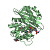


 PDBj
PDBj Assembly


 Mass: 96.063 Da / Num. of mol.: 3 / Source method: obtained synthetically / Formula: SO4
 Mass: 62.068 Da / Num. of mol.: 1 / Source method: obtained synthetically / Formula: C2H6O2
 Mass: 18.015 Da / Num. of mol.: 246 / Source method: isolated from a natural source / Formula: H2O
 Sample preparation
 / Beamline: X29A / Wavelength: 0.979 Å
 Processing