[English] 日本語
Yorodumi
- PDB-4q9a: Crystal structure of a putative GDSL-like lipase (PARMER_00689) f... -

+
Open data


ID or keywords:

Loading...

-
Basic information

Entry
Database: PDB / ID: 4q9a
TitleCrystal structure of a putative GDSL-like lipase (PARMER_00689) from Parabacteroides merdae ATCC 43184 at 2.86 A resolution
ComponentsTat pathway signal sequence domain protein
KeywordsHYDROLASE / Acylhydrolase family / PF13472 / Structural Genomics / Joint Center for Structural Genomics / JCSG / Protein Structure Initiative / PSI-BIOLOGY
Function / homology
Function and homology information


SGNH hydrolase / SGNH hydrolase-type esterase domain / GDSL-like Lipase/Acylhydrolase family / SGNH hydrolase superfamily / Twin arginine translocation (Tat) signal profile. / Twin-arginine translocation pathway, signal sequence / Rossmann fold / 3-Layer(aba) Sandwich / Alpha Beta
Similarity search - Domain/homology
Tat pathway signal sequence domain protein
Similarity search - Component
Biological speciesParabacteroides merdae (bacteria)
MethodX-RAY DIFFRACTION / SYNCHROTRON / SAD / Resolution: 2.86 Å
AuthorsJoint Center for Structural Genomics (JCSG)
CitationJournal: To be published
Title: Crystal structure of a putative GDSL-like lipase (PARMER_00689) from Parabacteroides merdae ATCC 43184 at 2.86 A resolution
Authors: Joint Center for Structural Genomics (JCSG)
History
DepositionApr 30, 2014Deposition site: RCSB / Processing site: RCSB
Revision 1.0Jun 25, 2014Provider: repository / Type: Initial release
Revision 1.1Nov 22, 2017Group: Refinement description / Category: software
Revision 1.2Feb 1, 2023Group: Database references / Derived calculations / Category: database_2 / struct_conn / struct_ref_seq_dif
Item: _database_2.pdbx_DOI / _database_2.pdbx_database_accession ..._database_2.pdbx_DOI / _database_2.pdbx_database_accession / _struct_conn.pdbx_leaving_atom_flag / _struct_ref_seq_dif.details

-
Structure visualization

Structure viewerMolecule:
MolmilJmol/JSmol

Downloads & links

-
Assembly

Deposited unit
A: Tat pathway signal sequence domain protein
B: Tat pathway signal sequence domain protein


Theoretical massNumber of molelcules
Total (without water)52,8042
Polymers52,8042
Non-polymers00
Water00
1


  • Idetical with deposited unit
  • defined by author&software
TypeNameSymmetry operationNumber
identity operation1_555x,y,z1
Buried area2080 Å2
ΔGint-18 kcal/mol
Surface area17780 Å2
MethodPISA
Unit cell
Length a, b, c (Å)128.829, 128.829, 149.650
Angle α, β, γ (deg.)90.000, 90.000, 90.000
Int Tables number97
Space group name H-MI422

-
Components

#1: Protein Tat pathway signal sequence domain protein


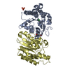
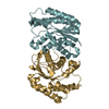
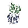
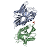
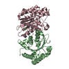
Mass: 26402.139 Da / Num. of mol.: 2 / Fragment: UNP residues 29-256
Source method: isolated from a genetically manipulated source
Source: (gene. exp.) Parabacteroides merdae (bacteria) / Gene: PARMER_00689 / Plasmid: SpeedET / Production host: Escherichia coli (E. coli) / Strain (production host): PB1 / References: UniProt: A7ABD4
Sequence detailsTHE CONSTRUCT (29-256) WAS EXPRESSED WITH A PURIFICATION TAG MGSDKIHHHHHHENLYFQG. THE TAG WAS ...THE CONSTRUCT (29-256) WAS EXPRESSED WITH A PURIFICATION TAG MGSDKIHHHHHHENLYFQG. THE TAG WAS REMOVED WITH TEV PROTEASE LEAVING ONLY A GLYCINE (0) FOLLOWED BY THE TARGET SEQUENCE.

-
Experimental details

-
Experiment

ExperimentMethod: X-RAY DIFFRACTION / Number of used crystals: 1

-
Sample preparation

CrystalDensity Matthews: 2.94 Å3/Da / Density % sol: 58.16 %
Crystal growTemperature: 277 K / Method: vapor diffusion, sitting drop / pH: 6.2
Details: 0.200M sodium chloride, 40.00% polyethylene glycol 400, 0.1M Na/K phosphate pH 6.2, NANODROP, VAPOR DIFFUSION, SITTING DROP, temperature 277K

-
Data collection

DiffractionMean temperature: 100 K
Diffraction sourceSource: SYNCHROTRON / Site: SSRL / Beamline: BL11-1 / Wavelength: 0.97891
DetectorType: DECTRIS PILATUS 6M / Detector: PIXEL / Date: Nov 27, 2013
Details: Flat mirror (vertical focusing); single crystal Si(111) bent monochromator (horizontal focusing)
RadiationMonochromator: single crystal Si(111) bent / Protocol: SINGLE WAVELENGTH / Monochromatic (M) / Laue (L): M / Scattering type: x-ray
Radiation wavelengthWavelength: 0.97891 Å / Relative weight: 1
ReflectionResolution: 2.86→48.817 Å / Num. obs: 14845 / % possible obs: 99.5 % / Observed criterion σ(I): -3 / Biso Wilson estimate: 68.432 Å2 / Rmerge(I) obs: 0.14 / Net I/σ(I): 11.62
Reflection shell
Resolution (Å)Rmerge(I) obsMean I/σ(I) obsNum. measured obsNum. unique obsDiffraction-ID% possible all
2.86-2.960.7632.294161417199.9
2.96-3.080.6252.695161467199.2
3.08-3.220.4713.590521452199.5
3.22-3.390.2855.7101621485199.9
3.39-3.60.1998.197181439199.6
3.6-3.880.1411.399581497199.9
3.88-4.260.11213.789331440199.2
4.26-4.870.08118.7101711492199.9
4.87-6.110.08118.994741514199.7
6.11-48.8170.05328.7100511636198.9

-
Phasing

PhasingMethod: SAD

-
Processing

Software
NameVersionClassificationNB
MolProbity3beta29model building
PDB_EXTRACT3.1data extraction
SHELXphasing
SHARPphasing
XSCALEdata scaling
BUSTER-TNT2.10.0refinement
XDSdata reduction
SHELXDphasing
BUSTER2.10.0refinement
RefinementMethod to determine structure: SAD / Resolution: 2.86→48.817 Å / Cor.coef. Fo:Fc: 0.9295 / Cor.coef. Fo:Fc free: 0.8958 / Occupancy max: 1 / Occupancy min: 0.75 / Cross valid method: THROUGHOUT / σ(F): 0
Details: 1. A MET-INHIBITION PROTOCOL WAS USED FOR SELENOMETHIONINE INCORPORATION DURING PROTEIN EXPRESSION. THE OCCUPANCY OF THE SE ATOMS IN THE MSE RESIDUES WAS REDUCED TO 0.75 FOR THE REDUCED ...Details: 1. A MET-INHIBITION PROTOCOL WAS USED FOR SELENOMETHIONINE INCORPORATION DURING PROTEIN EXPRESSION. THE OCCUPANCY OF THE SE ATOMS IN THE MSE RESIDUES WAS REDUCED TO 0.75 FOR THE REDUCED SCATTERING POWER DUE TO PARTIAL S-MET INCORPORATION. 2. ATOM RECORD CONTAINS SUM OF TLS AND RESIDUAL B FACTORS. ANISOU RECORD CONTAINS SUM OF TLS AND RESIDUAL U FACTORS. 3. THE EXPERIMENTAL PHASES WERE USED AS RESTRAINTS DURING REFINEMENT. 4. NCS RESTRAINTS WERE APPLIED USING BUSTER'S LSSR RESTRAINT REPRESENTATION (-AUTONCS). 5. RESIDUE SER55 IN BOTH CHAINS AS BEEN MODELED AS A PHOSPHORYLATED SERINE (SEP) BASED ON ELECTRON DENSITY.
RfactorNum. reflection% reflectionSelection details
Rfree0.2323 751 5.07 %RANDOM
Rwork0.1823 ---
obs0.1847 14826 99.67 %-
Displacement parametersBiso max: 116.75 Å2 / Biso mean: 52.8945 Å2 / Biso min: 28.44 Å2
Baniso -1Baniso -2Baniso -3
1-9.1979 Å20 Å20 Å2
2--9.1979 Å20 Å2
3----18.3959 Å2
Refine analyzeLuzzati coordinate error obs: 0.364 Å
Refinement stepCycle: LAST / Resolution: 2.86→48.817 Å
ProteinNucleic acidLigandSolventTotal
Num. atoms3509 0 0 0 3509
Refine LS restraints
Refine-IDTypeNumberRestraint functionWeightDev ideal
X-RAY DIFFRACTIONt_dihedral_angle_d1644SINUSOIDAL2
X-RAY DIFFRACTIONt_trig_c_planes98HARMONIC2
X-RAY DIFFRACTIONt_gen_planes515HARMONIC5
X-RAY DIFFRACTIONt_it3593HARMONIC20
X-RAY DIFFRACTIONt_nbd
X-RAY DIFFRACTIONt_improper_torsion
X-RAY DIFFRACTIONt_pseud_angle
X-RAY DIFFRACTIONt_chiral_improper_torsion455SEMIHARMONIC5
X-RAY DIFFRACTIONt_sum_occupancies
X-RAY DIFFRACTIONt_utility_distance
X-RAY DIFFRACTIONt_utility_angle
X-RAY DIFFRACTIONt_utility_torsion
X-RAY DIFFRACTIONt_ideal_dist_contact4106SEMIHARMONIC4
X-RAY DIFFRACTIONt_bond_d3593HARMONIC20.01
X-RAY DIFFRACTIONt_angle_deg4872HARMONIC21.06
X-RAY DIFFRACTIONt_omega_torsion3.11
X-RAY DIFFRACTIONt_other_torsion3.08
LS refinement shellResolution: 2.86→3.09 Å / Total num. of bins used: 7
RfactorNum. reflection% reflection
Rfree0.2874 171 5.71 %
Rwork0.225 2824 -
all0.2285 2995 -
obs--99.67 %
Refinement TLS params.

Method: refined / Refine-ID: X-RAY DIFFRACTION

IDL112)L122)L132)L222)L232)L332)S11 (Å °)S12 (Å °)S13 (Å °)S21 (Å °)S22 (Å °)S23 (Å °)S31 (Å °)S32 (Å °)S33 (Å °)T112)T122)T132)T222)T232)T332)Origin x (Å)Origin y (Å)Origin z (Å)
11.6275-0.37130.5891.6018-0.33181.49090.05840.1636-0.0625-0.21920.0120.18150.0775-0.1048-0.0704-0.1447-0.0213-0.0451-0.14590.02190.170524.80247.347915.4288
21.03770.36340.57730.9849-0.06651.4146-0.0018-0.09150.0950.15650.0070.07090.02210.013-0.0052-0.14140.02230.0215-0.12880.0140.200935.321173.057635.1408
Refinement TLS group
IDRefine-IDRefine TLS-IDSelection detailsAuth asym-IDAuth seq-ID
1X-RAY DIFFRACTION1{ A|38 - 256 }A38 - 256
2X-RAY DIFFRACTION2{ B|39 - 256 }B39 - 256

+
About Yorodumi

-
News

-
Feb 9, 2022. New format data for meta-information of EMDB entries

New format data for meta-information of EMDB entries

  • Version 3 of the EMDB header file is now the official format.
  • The previous official version 1.9 will be removed from the archive.

Related info.:EMDB header

External links:wwPDB to switch to version 3 of the EMDB data model

-
Aug 12, 2020. Covid-19 info

Covid-19 info

URL: https://pdbjlvh1.pdbj.org/emnavi/covid19.php

New page: Covid-19 featured information page in EM Navigator.

Related info.:Covid-19 info / Mar 5, 2020. Novel coronavirus structure data

+
Mar 5, 2020. Novel coronavirus structure data

Novel coronavirus structure data

Related info.:Yorodumi Speices / Aug 12, 2020. Covid-19 info

External links:COVID-19 featured content - PDBj / Molecule of the Month (242):Coronavirus Proteases

+
Jan 31, 2019. EMDB accession codes are about to change! (news from PDBe EMDB page)

EMDB accession codes are about to change! (news from PDBe EMDB page)

  • The allocation of 4 digits for EMDB accession codes will soon come to an end. Whilst these codes will remain in use, new EMDB accession codes will include an additional digit and will expand incrementally as the available range of codes is exhausted. The current 4-digit format prefixed with “EMD-” (i.e. EMD-XXXX) will advance to a 5-digit format (i.e. EMD-XXXXX), and so on. It is currently estimated that the 4-digit codes will be depleted around Spring 2019, at which point the 5-digit format will come into force.
  • The EM Navigator/Yorodumi systems omit the EMD- prefix.

Related info.:Q: What is EMD? / ID/Accession-code notation in Yorodumi/EM Navigator

External links:EMDB Accession Codes are Changing Soon! / Contact to PDBj

+
Jul 12, 2017. Major update of PDB

Major update of PDB

  • wwPDB released updated PDB data conforming to the new PDBx/mmCIF dictionary.
  • This is a major update changing the version number from 4 to 5, and with Remediation, in which all the entries are updated.
  • In this update, many items about electron microscopy experimental information are reorganized (e.g. em_software).
  • Now, EM Navigator and Yorodumi are based on the updated data.

External links:wwPDB Remediation / Enriched Model Files Conforming to OneDep Data Standards Now Available in the PDB FTP Archive

-
Yorodumi

Thousand views of thousand structures

  • Yorodumi is a browser for structure data from EMDB, PDB, SASBDB, etc.
  • This page is also the successor to EM Navigator detail page, and also detail information page/front-end page for Omokage search.
  • The word "yorodu" (or yorozu) is an old Japanese word meaning "ten thousand". "mi" (miru) is to see.

Related info.:EMDB / PDB / SASBDB / Comparison of 3 databanks / Yorodumi Search / Aug 31, 2016. New EM Navigator & Yorodumi / Yorodumi Papers / Jmol/JSmol / Function and homology information / Changes in new EM Navigator and Yorodumi

Read more