- PDB-4q5t: Crystal structure of an atmB (putative membrane lipoprotein) from... -
+
Open data
ID or keywords:
Loading...
-
Basic information
Entry
Database: PDB / ID: 4q5t
Title
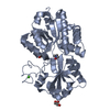
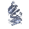
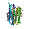
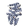
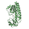
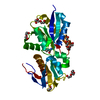
Crystal structure of an atmB (putative membrane lipoprotein) from Streptococcus mutans UA159 at 1.91 A resolution
Components
Lipoprotein
Keywords
TRANSPORT PROTEIN / methionine-binding / NLPA lipoprotein / PF03180 family / Structural Genomics / Joint Center for Structural Genomics / JCSG / Protein Structure Initiative / PSI-BIOLOGY
Mass: 18.015 Da / Num. of mol.: 154 / Source method: isolated from a natural source / Formula: H2O
Has protein modification
Y
Sequence details
THIS CONSTRUCT WAS EXPRESSED WITH AN N-TERMINAL PURIFICATION TAG MGSDKIHHHHHHENLYFQG. THE TAG WAS ...THIS CONSTRUCT WAS EXPRESSED WITH AN N-TERMINAL PURIFICATION TAG MGSDKIHHHHHHENLYFQG. THE TAG WAS REMOVED WITH TEV PROTEASE LEAVING ONLY A GLYCINE (0) FOLLOWED BY RESIDUES 25-280 OF THE TARGET SEQUENCE.
-
Experimental details
-
Experiment
Experiment
Method: X-RAY DIFFRACTION / Number of used crystals: 1
-
Sample preparation
Crystal
Density Matthews: 2.54 Å3/Da / Density % sol: 51.6 %
Resolution: 1.91→28.777 Å / Num. obs: 23167 / % possible obs: 98 % / Observed criterion σ(I): -3 / Biso Wilson estimate: 23.682 Å2 / Rmerge(I) obs: 0.057 / Net I/σ(I): 11.11
Reflection shell
Resolution (Å)
Rmerge(I) obs
Mean I/σ(I) obs
Num. measured obs
Num. unique obs
Diffraction-ID
% possible all
1.91-1.98
0.448
2
10172
4385
1
96.6
1.98-2.06
0.31
2.7
10262
4401
1
99
2.06-2.15
0.243
3.6
9478
4093
1
96.4
2.15-2.26
0.168
5.2
9817
4212
1
97.7
2.26-2.41
0.132
6.2
10937
4652
1
98.9
2.41-2.59
0.098
8
9984
4228
1
99.2
2.59-2.85
0.075
10.6
10243
4367
1
98.5
2.85-3.26
0.049
15.4
10277
4370
1
99.2
3.26-4.1
0.031
24.8
9897
4280
1
96.2
4.1-28.777
0.022
32.3
10512
4406
1
98.1
-
Phasing
Phasing
Method: MAD
-
Processing
Software
Name
Version
Classification
NB
MolProbity
3beta29
modelbuilding
PDB_EXTRACT
3.1
dataextraction
SHELX
phasing
SHARP
phasing
XSCALE
March15, 2012
datascaling
PHENIX
1.8.2
refinement
XDS
datareduction
SHELXD
phasing
Refinement
Method to determine structure: MAD / Resolution: 1.907→28.777 Å / Occupancy max: 1 / Occupancy min: 0.5 / SU ML: 0.21 / σ(F): 1.22 / Phase error: 23.87 / Stereochemistry target values: MLHL Details: 1. ZERO OCCUPANCY HYDROGENS WERE INCLUDED DURING REFINEMENT TO IMPROVE THE ANTI-BUMPING RESTRAINTS. 2. ATOM RECORDS CONTAIN SUM OF TLS AND RESIDUAL B FACTORS. 3. ANISOU RECORDS CONTAIN SUM ...Details: 1. ZERO OCCUPANCY HYDROGENS WERE INCLUDED DURING REFINEMENT TO IMPROVE THE ANTI-BUMPING RESTRAINTS. 2. ATOM RECORDS CONTAIN SUM OF TLS AND RESIDUAL B FACTORS. 3. ANISOU RECORDS CONTAIN SUM OF TLS AND RESIDUAL U FACTORS. 4. A MET-INHIBITION PROTOCOL WAS USED FOR SELENOMETHIONINE INCORPORATION DURING PROTEIN EXPRESSION. THE OCCUPANCY OF THE SE ATOMS IN THE MSE RESIDUES WAS REDUCED TO 0.75 FOR THE REDUCED SCATTERING POWER DUE TO PARTIAL S-MET INCORPORATION. 5. LIGAND SELENOMETHIONINE HAS BEEN MODELED BASED ON DENSITY AND ANOMALOUS DIFFERENCE FOURIER MAP. 6. NONAETHYLENE GLYCOL (2PE) MOLECULES FROM THE CRYSTALLIZATION SOLUTION ARE MODELED.
Rfactor
Num. reflection
% reflection
Rfree
0.2302
1189
5.14 %
Rwork
0.1827
-
-
obs
0.185
23128
99.24 %
Solvent computation
Shrinkage radii: 0.9 Å / VDW probe radii: 1.11 Å / Solvent model: FLAT BULK SOLVENT MODEL
In the structure databanks used in Yorodumi, some data are registered as the other names, "COVID-19 virus" and "2019-nCoV". Here are the details of the virus and the list of structure data.
Jan 31, 2019. EMDB accession codes are about to change! (news from PDBe EMDB page)
EMDB accession codes are about to change! (news from PDBe EMDB page)
The allocation of 4 digits for EMDB accession codes will soon come to an end. Whilst these codes will remain in use, new EMDB accession codes will include an additional digit and will expand incrementally as the available range of codes is exhausted. The current 4-digit format prefixed with “EMD-” (i.e. EMD-XXXX) will advance to a 5-digit format (i.e. EMD-XXXXX), and so on. It is currently estimated that the 4-digit codes will be depleted around Spring 2019, at which point the 5-digit format will come into force.
The EM Navigator/Yorodumi systems omit the EMD- prefix.
Related info.:Q: What is EMD? / ID/Accession-code notation in Yorodumi/EM Navigator
Yorodumi is a browser for structure data from EMDB, PDB, SASBDB, etc.
This page is also the successor to EM Navigator detail page, and also detail information page/front-end page for Omokage search.
The word "yorodu" (or yorozu) is an old Japanese word meaning "ten thousand". "mi" (miru) is to see.
Related info.:EMDB / PDB / SASBDB / Comparison of 3 databanks / Yorodumi Search / Aug 31, 2016. New EM Navigator & Yorodumi / Yorodumi Papers / Jmol/JSmol / Function and homology information / Changes in new EM Navigator and Yorodumi
 Movie
Movie Controller

 Yorodumi
 Open data
 Basic information
 Components
 Keywords
 Function and homology information
 Streptococcus mutans UA159 (bacteria)
 X-RAY DIFFRACTION /
  Authors
 Citation
 Structure visualization
 Downloads & links
 Other downloads
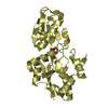


 PDBj
PDBj


 Assembly
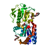

 Type: L-peptide linking / Mass: 196.106 Da / Num. of mol.: 1 / Source method: obtained synthetically / Formula: C5H11NO2Se
 Mass: 414.488 Da / Num. of mol.: 3 / Source method: obtained synthetically / Formula: C18H38O10 / Comment: precipitant*YM
 Mass: 18.015 Da / Num. of mol.: 154 / Source method: isolated from a natural source / Formula: H2O
 Sample preparation
 / Beamline: 8.2.2 / Wavelength: 0.979068,0.979338,0.953725
 Processing