 Movie
Movie Controller


+ Open data

- Basic information
| Entry | Database: PDB / ID: 4q4f | |||||||||
|---|---|---|---|---|---|---|---|---|---|---|
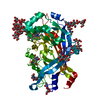
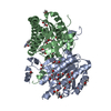
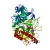
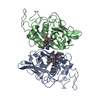
| Title | Crystal structure of LIMP-2 (space group C2) | |||||||||
 Components Components | Lysosome membrane protein 2 | |||||||||
 Keywords Keywords | CELL ADHESION / Lysosomal targeting / beta-glucocerebrosidase / CI-MPR / ER / endosome / lysosome | |||||||||
| Function / homology |  Function and homology information Function and homology informationregulation of glucosylceramide catabolic process / regulation of carbohydrate catabolic process / regulation of endosome organization / aminophospholipid transport / regulation of lysosome organization / protein targeting to lysosome / phosphatidylcholine binding / scavenger receptor activity / cargo receptor activity / cholesterol binding ...regulation of glucosylceramide catabolic process / regulation of carbohydrate catabolic process / regulation of endosome organization / aminophospholipid transport / regulation of lysosome organization / protein targeting to lysosome / phosphatidylcholine binding / scavenger receptor activity / cargo receptor activity / cholesterol binding / phosphatidylserine binding / lysosomal lumen / receptor-mediated endocytosis / positive regulation of neuron projection development / clathrin-coated endocytic vesicle membrane / autophagy / endocytic vesicle membrane / transmembrane signaling receptor activity / late endosome membrane / Cargo recognition for clathrin-mediated endocytosis / Clathrin-mediated endocytosis / protein-folding chaperone binding / virus receptor activity / endosome membrane / Golgi membrane / lysosomal membrane / focal adhesion / endoplasmic reticulum membrane / enzyme binding / protein homodimerization activity / extracellular exosome / membrane / plasma membrane Similarity search - Function | |||||||||
| Biological species |  Homo sapiens (human) Homo sapiens (human) | |||||||||
| Method |  X-RAY DIFFRACTION / SYNCHROTRON / MOLECULAR REPLACEMENT / Resolution: 2.8 Å X-RAY DIFFRACTION / SYNCHROTRON / MOLECULAR REPLACEMENT / Resolution: 2.8 Å | |||||||||
 Authors Authors | Zhao, Y. / Ren, J. / Padilla-Parra, S. / Fry, L.E. / Stuart, D.I. | |||||||||
 Citation Citation | Journal: Nat Commun / Year: 2014 Title: Lysosome sorting of beta-glucocerebrosidase by LIMP-2 is targeted by the mannose 6-phosphate receptor. Authors: Zhao, Y. / Ren, J. / Padilla-Parra, S. / Fry, E.E. / Stuart, D.I. | |||||||||
| History |
|
- Structure visualization
| Structure viewer | Molecule: MolmilJmol/JSmol |
|---|
- Downloads & links
-Download
| PDBx/mmCIF format | 4q4f.cif.gz | 193.6 KB | Display | PDBx/mmCIF format |
|---|---|---|---|---|
| PDB format | pdb4q4f.ent.gz | 155.8 KB | Display | PDB format |
| PDBx/mmJSON format | 4q4f.json.gz | Tree view | PDBx/mmJSON format | |
| Others |  Other downloads Other downloads |
-Validation report
| Arichive directory | https://data.pdbj.org/pub/pdb/validation_reports/q4/4q4fftp://data.pdbj.org/pub/pdb/validation_reports/q4/4q4f | HTTPS FTP |
|---|
-Related structure data
| Related structure data |  4q4bSC S: Starting model for refinement C: citing same article ( |
|---|---|
| Similar structure data |
-Links
 PDBj
PDBj- Assembly
| Deposited unit | 
| ||||||||
|---|---|---|---|---|---|---|---|---|---|
| 1 |
| ||||||||
| Unit cell |
|
-Components
-Protein / Non-polymers , 2 types, 2 molecules A
| #1: Protein | Mass: 47727.750 Da / Num. of mol.: 1 / Fragment: Human Limp-2 luminal domain Source method: isolated from a genetically manipulated source Source: (gene. exp.) Homo sapiens (human) / Gene: SCARB2, CD36L2, LIMP2, LIMPII / Plasmid: pNeoSec / Cell line (production host): HEK 293S GNTI- / Production host: homo sapiens (human) / References: UniProt: Q14108 |
|---|---|
| #8: Chemical | ChemComp-CL /  Mass: 35.453 Da / Num. of mol.: 1 / Source method: obtained synthetically / Formula: Cl Mass: 35.453 Da / Num. of mol.: 1 / Source method: obtained synthetically / Formula: Cl |
-Sugars , 6 types, 8 molecules 
| #2: Polysaccharide | Source method: isolated from a genetically manipulated source #3: Polysaccharide | alpha-D-mannopyranose-(1-3)-alpha-D-mannopyranose-(1-6)-[alpha-D-mannopyranose-(1-3)]beta-D- ...alpha-D-mannopyranose-(1-3)-alpha-D-mannopyranose-(1-6)-[alpha-D-mannopyranose-(1-3)]beta-D-mannopyranose-(1-4)-2-acetamido-2-deoxy-beta-D-glucopyranose-(1-4)-2-acetamido-2-deoxy-beta-D-glucopyranose | Source method: isolated from a genetically manipulated source #4: Polysaccharide | beta-D-mannopyranose-(1-4)-2-acetamido-2-deoxy-beta-D-glucopyranose-(1-4)-2-acetamido-2-deoxy-beta- ...beta-D-mannopyranose-(1-4)-2-acetamido-2-deoxy-beta-D-glucopyranose-(1-4)-2-acetamido-2-deoxy-beta-D-glucopyranose | Source method: isolated from a genetically manipulated source #5: Polysaccharide | 2-acetamido-2-deoxy-beta-D-glucopyranose-(1-4)-2-acetamido-2-deoxy-beta-D-glucopyranose | Source method: isolated from a genetically manipulated source #6: Polysaccharide | 6-O-phosphono-beta-D-mannopyranose-(1-2)-alpha-D-mannopyranose-(1-6)-alpha-D-mannopyranose-(1-6)- ...6-O-phosphono-beta-D-mannopyranose-(1-2)-alpha-D-mannopyranose-(1-6)-alpha-D-mannopyranose-(1-6)-[alpha-D-mannopyranose-(1-3)]beta-D-mannopyranose-(1-4)-2-acetamido-2-deoxy-beta-D-glucopyranose-(1-4)-2-acetamido-2-deoxy-beta-D-glucopyranose | Source method: isolated from a genetically manipulated source #7: Sugar |  Type: D-saccharide, beta linking / Mass: 221.208 Da / Num. of mol.: 2 Type: D-saccharide, beta linking / Mass: 221.208 Da / Num. of mol.: 2Source method: isolated from a genetically manipulated source Formula: C8H15NO6 |
|---|
-Details
| Has protein modification | Y |
|---|
-Experimental details
-Experiment
| Experiment | Method: X-RAY DIFFRACTION / Number of used crystals: 1 |
|---|
- Sample preparation
| Crystal | Density Matthews: 3.33 Å3/Da / Density % sol: 63.04 % |
|---|---|
| Crystal grow | Temperature: 294 K / Method: vapor diffusion, sitting drop / pH: 6.5 Details: 30 %w/v Polyethylene Glycol 8000, 0.2 M Ammonium Sulphate, 0.1 M Sodium Cacodylate, pH 6.5, VAPOR DIFFUSION, SITTING DROP, temperature 294K |
-Data collection
| Diffraction | Mean temperature: 100 K |
|---|---|
| Diffraction source | Source: SYNCHROTRON / Site: Diamond  / Beamline: I24 / Wavelength: 0.9686 Å / Beamline: I24 / Wavelength: 0.9686 Å |
| Detector | Type: PSI PILATUS 6M / Detector: PIXEL / Date: Jul 5, 2012 |
| Radiation | Monochromator: double crystal monochromator / Protocol: SINGLE WAVELENGTH / Monochromatic (M) / Laue (L): M / Scattering type: x-ray |
| Radiation wavelength | Wavelength: 0.9686 Å / Relative weight: 1 |
| Reflection | Resolution: 2.8→50 Å / Num. obs: 15244 / % possible obs: 96.2 % / Observed criterion σ(I): -3 / Redundancy: 2.5 % / Rmerge(I) obs: 0.119 / Net I/σ(I): 7.1 |
| Reflection shell | Resolution: 2.8→2.9 Å / Redundancy: 2.1 % / Rmerge(I) obs: 0.378 / Mean I/σ(I) obs: 1.8 / % possible all: 91.5 |
- Processing
| Software |
| ||||||||||||||||||||||||||||||||||||||||||||||||||||||||||||||||||||||||||||||||||||||||||||||||||||||||||||||||||||||||||||||||||||||||||||||||||||||||||||||||||||||||||||||||||||||
|---|---|---|---|---|---|---|---|---|---|---|---|---|---|---|---|---|---|---|---|---|---|---|---|---|---|---|---|---|---|---|---|---|---|---|---|---|---|---|---|---|---|---|---|---|---|---|---|---|---|---|---|---|---|---|---|---|---|---|---|---|---|---|---|---|---|---|---|---|---|---|---|---|---|---|---|---|---|---|---|---|---|---|---|---|---|---|---|---|---|---|---|---|---|---|---|---|---|---|---|---|---|---|---|---|---|---|---|---|---|---|---|---|---|---|---|---|---|---|---|---|---|---|---|---|---|---|---|---|---|---|---|---|---|---|---|---|---|---|---|---|---|---|---|---|---|---|---|---|---|---|---|---|---|---|---|---|---|---|---|---|---|---|---|---|---|---|---|---|---|---|---|---|---|---|---|---|---|---|---|---|---|---|---|
| Refinement | Method to determine structure: MOLECULAR REPLACEMENT Starting model: pdb entry 4Q4B Resolution: 2.8→50 Å / Cor.coef. Fo:Fc: 0.931 / Cor.coef. Fo:Fc free: 0.885 / SU B: 46.978 / SU ML: 0.386 / Cross valid method: THROUGHOUT / ESU R: 2.797 / ESU R Free: 0.385 / Stereochemistry target values: MAXIMUM LIKELIHOOD / Details: HYDROGENS HAVE BEEN ADDED IN THE RIDING POSITIONS
| ||||||||||||||||||||||||||||||||||||||||||||||||||||||||||||||||||||||||||||||||||||||||||||||||||||||||||||||||||||||||||||||||||||||||||||||||||||||||||||||||||||||||||||||||||||||
| Solvent computation | Ion probe radii: 0.8 Å / Shrinkage radii: 0.8 Å / VDW probe radii: 1.2 Å / Solvent model: MASK | ||||||||||||||||||||||||||||||||||||||||||||||||||||||||||||||||||||||||||||||||||||||||||||||||||||||||||||||||||||||||||||||||||||||||||||||||||||||||||||||||||||||||||||||||||||||
| Displacement parameters | Biso mean: 71.184 Å2
| ||||||||||||||||||||||||||||||||||||||||||||||||||||||||||||||||||||||||||||||||||||||||||||||||||||||||||||||||||||||||||||||||||||||||||||||||||||||||||||||||||||||||||||||||||||||
| Refinement step | Cycle: LAST / Resolution: 2.8→50 Å
| ||||||||||||||||||||||||||||||||||||||||||||||||||||||||||||||||||||||||||||||||||||||||||||||||||||||||||||||||||||||||||||||||||||||||||||||||||||||||||||||||||||||||||||||||||||||
| Refine LS restraints |
|