 Movie
Movie Controller

[English] 日本語
 Yorodumi
- PDB-1the: CRYSTAL STRUCTURES OF RECOMBINANT RAT CATHEPSIN B AND A CATHEPSIN... -
+ Open data

- Basic information
| Entry | Database: PDB / ID: 1the | ||||||
|---|---|---|---|---|---|---|---|
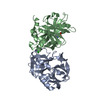
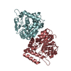
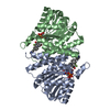
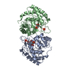
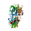
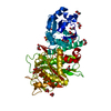
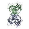
| Title | CRYSTAL STRUCTURES OF RECOMBINANT RAT CATHEPSIN B AND A CATHEPSIN B-INHIBITOR COMPLEX: IMPLICATIONS FOR STRUCTURE-BASED INHIBITOR DESIGN | ||||||
 Components Components | Cathepsin B | ||||||
 Keywords Keywords | HYDROLASE/HYDROLASE INHIBITOR / THIOL PROTEASE / Glycoprotein / Protease / HYDROLASE-HYDROLASE INHIBITOR complex | ||||||
| Function / homology |  Function and homology information Function and homology informationTrafficking and processing of endosomal TLR / Assembly of collagen fibrils and other multimeric structures / Collagen degradation / kininogen binding / Degradation of CDH1 / cathepsin B / response to interleukin-4 / peptidase inhibitor complex / MHC class II antigen presentation / response to kainic acid ...Trafficking and processing of endosomal TLR / Assembly of collagen fibrils and other multimeric structures / Collagen degradation / kininogen binding / Degradation of CDH1 / cathepsin B / response to interleukin-4 / peptidase inhibitor complex / MHC class II antigen presentation / response to kainic acid / thyroid hormone generation / cellular response to thyroid hormone stimulus / proteoglycan binding / response to amine / Neutrophil degranulation / response to dexamethasone / decidualization / collagen catabolic process / response to mechanical stimulus / response to glucose / skeletal muscle tissue development / collagen binding / peptide binding / epithelial cell differentiation / cysteine-type peptidase activity / response to cytokine / : / protein catabolic process / cellular response to mechanical stimulus / sarcolemma / caveola / response to peptide hormone / autophagy / melanosome / peptidase activity / neuron apoptotic process / endopeptidase activity / spermatogenesis / response to ethanol / lysosome / apical plasma membrane / external side of plasma membrane / cysteine-type endopeptidase activity / symbiont entry into host cell / protein-containing complex binding / perinuclear region of cytoplasm / cell surface / proteolysis / : / extracellular region / identical protein binding Similarity search - Function | ||||||
| Biological species |  | ||||||
| Method |  X-RAY DIFFRACTION / Resolution: 1.9 Å X-RAY DIFFRACTION / Resolution: 1.9 Å | ||||||
 Authors Authors | Huber, C.P. / Jia, Z. | ||||||
 Citation Citation | Journal: J.Biol.Chem. / Year: 1995 Title: Crystal structures of recombinant rat cathepsin B and a cathepsin B-inhibitor complex. Implications for structure-based inhibitor design. Authors: Jia, Z. / Hasnain, S. / Hirama, T. / Lee, X. / Mort, J.S. / To, R. / Huber, C.P. #1: Journal: J.Biol.Chem. / Year: 1990Title: Crystallization of Recombinant Rat Cathepsin B Authors: Lee, X. / Ahmed, F.R. / Hirama, T. / Huber, C.P. / Rose, D.R. / To, R. / Hasnain, S. / Tam, A. / Mort, J.S. | ||||||
| History |
| ||||||
| Remark 700 | SHEET THERE IS A BIFURCATED SHEET IN EACH CHAIN OF THIS ENTRY. A BIFURCATED SHEET IS PRESENTED AS ...SHEET THERE IS A BIFURCATED SHEET IN EACH CHAIN OF THIS ENTRY. A BIFURCATED SHEET IS PRESENTED AS TWO SHEETS WITH ONE OR MORE STRANDS IN COMMON. |
- Structure visualization
| Structure viewer | Molecule: MolmilJmol/JSmol |
|---|
- Downloads & links
-Download
| PDBx/mmCIF format | 1the.cif.gz | 116.8 KB | Display | PDBx/mmCIF format |
|---|---|---|---|---|
| PDB format | pdb1the.ent.gz | 89.5 KB | Display | PDB format |
| PDBx/mmJSON format | 1the.json.gz | Tree view | PDBx/mmJSON format | |
| Others |  Other downloads Other downloads |
-Validation report
| Arichive directory | https://data.pdbj.org/pub/pdb/validation_reports/th/1theftp://data.pdbj.org/pub/pdb/validation_reports/th/1the | HTTPS FTP |
|---|
-Related structure data


-Links
 PDBj
PDBj


- Assembly
| Deposited unit | 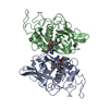
| ||||||||
|---|---|---|---|---|---|---|---|---|---|
| 1 | 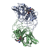
| ||||||||
| Unit cell |
| ||||||||
| Noncrystallographic symmetry (NCS) | NCS oper: (Code: given Matrix: (0.6405, 0.7349, 0.2229), Vector: |
-Components
| #1: Protein | Mass: 28441.562 Da / Num. of mol.: 2 / Mutation: S115A Source method: isolated from a genetically manipulated source Details: RECOMBINANT RAT ENZYME / Source: (gene. exp.)  #2: Chemical |   Type: peptide-like, Peptide-like / Class: Inhibitor / Mass: 519.013 Da / Num. of mol.: 2 / Source method: obtained synthetically / Formula: C25H33ClN5O5 Type: peptide-like, Peptide-like / Class: Inhibitor / Mass: 519.013 Da / Num. of mol.: 2 / Source method: obtained synthetically / Formula: C25H33ClN5O5References: amino{[(4R)-4-{[(benzyloxy)carbonyl]amino}-5-({(1R)-1-[(benzyloxy)methyl]-3-chloro-2-oxopropyl}amino)-5- oxopentyl]amino}methaniminium #3: Water | ChemComp-HOH / |  Mass: 18.015 Da / Num. of mol.: 238 / Source method: isolated from a natural source / Formula: H2O Mass: 18.015 Da / Num. of mol.: 238 / Source method: isolated from a natural source / Formula: H2OHas protein modification | Y | Sequence details | THE VAL 223 ALA SUBSTITUTION WAS PRESENT IN THE PLASMID SUPPLIED TO DR. J. S. MORT BY DR. SHU CHAN ...THE VAL 223 ALA SUBSTITUTI | |
|---|
-Experimental details
-Experiment
| Experiment | Method: X-RAY DIFFRACTION |
|---|
- Sample preparation
| Crystal | Density Matthews: 2.31 Å3/Da / Density % sol: 46.67 % | ||||||||||||||||||||||||||||||||||||||||||||||||
|---|---|---|---|---|---|---|---|---|---|---|---|---|---|---|---|---|---|---|---|---|---|---|---|---|---|---|---|---|---|---|---|---|---|---|---|---|---|---|---|---|---|---|---|---|---|---|---|---|---|
| Crystal grow | *PLUS pH: 6 / Method: vapor diffusion, hanging drop | ||||||||||||||||||||||||||||||||||||||||||||||||
| Components of the solutions | *PLUS
|
-Data collection
| Diffraction source | Wavelength: 1.5418 Å |
|---|---|
| Detector | Type: XUONG-HAMLIN MULTIWIRE / Detector: AREA DETECTOR / Date: Sep 1, 1992 |
| Radiation | Monochromatic (M) / Laue (L): M / Scattering type: x-ray |
| Radiation wavelength | Wavelength: 1.5418 Å / Relative weight: 1 |
| Reflection | Resolution: 1.9→14.3 Å / Num. obs: 39749 / % possible obs: 97.7 % / Observed criterion σ(I): 0 / Redundancy: 4 % / Rmerge(I) obs: 0.07 |
- Processing
| Software |
| ||||||||||||||||||||||||||||||||||||||||||||||||||||||||||||
|---|---|---|---|---|---|---|---|---|---|---|---|---|---|---|---|---|---|---|---|---|---|---|---|---|---|---|---|---|---|---|---|---|---|---|---|---|---|---|---|---|---|---|---|---|---|---|---|---|---|---|---|---|---|---|---|---|---|---|---|---|---|
| Refinement | Resolution: 1.9→8 Å / σ(F): 2 Details: RESIDUE ASN 222 OF BOTH CHAINS HAS PHI-PSI ANGLES OUTSIDE THE NORMALLY PERMITTED RANGE BECAUSE ITS SIDECHAIN OXYGEN ATOM IS INVOLVED IN A HYDROGEN BOND WITH THE ADJACENT MAIN-CHAIN N-H 223.
| ||||||||||||||||||||||||||||||||||||||||||||||||||||||||||||
| Displacement parameters | Biso mean: 19.4 Å2 | ||||||||||||||||||||||||||||||||||||||||||||||||||||||||||||
| Refine analyze | Luzzati coordinate error obs: 0.19 Å | ||||||||||||||||||||||||||||||||||||||||||||||||||||||||||||
| Refinement step | Cycle: LAST / Resolution: 1.9→8 Å
| ||||||||||||||||||||||||||||||||||||||||||||||||||||||||||||
| Refine LS restraints |
|
