 Movie
Movie Controller

[English] 日本語
 Yorodumi
- PDB-4pw6: structure of UHRF2-SRA in complex with a 5hmC-containing DNA, com... -
+ Open data

- Basic information
| Entry | Database: PDB / ID: 4pw6 | ||||||
|---|---|---|---|---|---|---|---|
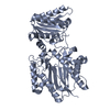
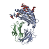
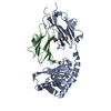
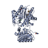
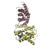
| Title | structure of UHRF2-SRA in complex with a 5hmC-containing DNA, complex II | ||||||
 Components Components |
| ||||||
 Keywords Keywords | LIGASE/DNA / SRA / 5hmC binding / 5hmC-containing DNA / hydroxymethylation / nuclear / LIGASE-DNA complex | ||||||
| Function / homology |  Function and homology information Function and homology informationSUMO transferase activity / histone H3K9me2/3 reader activity / negative regulation of gene expression via chromosomal CpG island methylation / protein sumoylation / pericentric heterochromatin / protein autoubiquitination / heterochromatin / SUMOylation of transcription cofactors / RING-type E3 ubiquitin transferase / ubiquitin-protein transferase activity ...SUMO transferase activity / histone H3K9me2/3 reader activity / negative regulation of gene expression via chromosomal CpG island methylation / protein sumoylation / pericentric heterochromatin / protein autoubiquitination / heterochromatin / SUMOylation of transcription cofactors / RING-type E3 ubiquitin transferase / ubiquitin-protein transferase activity / ubiquitin protein ligase activity / histone binding / RNA polymerase II-specific DNA-binding transcription factor binding / cell differentiation / regulation of cell cycle / protein ubiquitination / regulation of transcription by RNA polymerase II / DNA binding / zinc ion binding / nucleoplasm / nucleus Similarity search - Function | ||||||
| Biological species |  Homo sapiens (human) Homo sapiens (human) | ||||||
| Method |  X-RAY DIFFRACTION / SYNCHROTRON / MOLECULAR REPLACEMENT / Resolution: 3.789 Å X-RAY DIFFRACTION / SYNCHROTRON / MOLECULAR REPLACEMENT / Resolution: 3.789 Å | ||||||
 Authors Authors | Zhou, T. / Xiong, J. / Wang, M. / Yang, N. / Wong, J. / Zhu, B. / Xu, R.M. | ||||||
 Citation Citation | Journal: Mol.Cell / Year: 2014 Title: Structural Basis for Hydroxymethylcytosine Recognition by the SRA Domain of UHRF2. Authors: Zhou, T. / Xiong, J. / Wang, M. / Yang, N. / Wong, J. / Zhu, B. / Xu, R.M. | ||||||
| History |
|
- Structure visualization
| Structure viewer | Molecule: MolmilJmol/JSmol |
|---|
- Downloads & links
-Download
| PDBx/mmCIF format | 4pw6.cif.gz | 100.3 KB | Display | PDBx/mmCIF format |
|---|---|---|---|---|
| PDB format | pdb4pw6.ent.gz | 73.5 KB | Display | PDB format |
| PDBx/mmJSON format | 4pw6.json.gz | Tree view | PDBx/mmJSON format | |
| Others |  Other downloads Other downloads |
-Validation report
| Arichive directory | https://data.pdbj.org/pub/pdb/validation_reports/pw/4pw6ftp://data.pdbj.org/pub/pdb/validation_reports/pw/4pw6 | HTTPS FTP |
|---|
-Related structure data
| Related structure data |  4pw5C  4pw7C  3olnS C: citing same article ( S: Starting model for refinement |
|---|---|
| Similar structure data |


-Links
 PDBj
PDBj


- Assembly
| Deposited unit | 
| ||||||||
|---|---|---|---|---|---|---|---|---|---|
| 1 |
| ||||||||
| Unit cell |
|
-Components
| #1: Protein | Mass: 25579.746 Da / Num. of mol.: 2 Source method: isolated from a genetically manipulated source Source: (gene. exp.) Homo sapiens (human) / Gene: UHRF2, NIRF, RNF107 / Plasmid: pET28a-smt / Production host:  References: UniProt: Q96PU4, Ligases; Forming carbon-nitrogen bonds; Acid-amino-acid ligases (peptide synthases) #2: DNA chain | | Mass: 3747.465 Da / Num. of mol.: 1 / Source method: obtained synthetically #3: DNA chain | | Mass: 3606.381 Da / Num. of mol.: 1 / Source method: obtained synthetically |
|---|
-Experimental details
-Experiment
| Experiment | Method: X-RAY DIFFRACTION / Number of used crystals: 1 |
|---|
- Sample preparation
| Crystal | Density Matthews: 2.33 Å3/Da / Density % sol: 47.31 % |
|---|---|
| Crystal grow | Temperature: 289 K / Method: vapor diffusion, sitting drop / pH: 8 Details: 0.1 M Tris pH 8.0 and 8% PEG-8000, VAPOR DIFFUSION, SITTING DROP, temperature 289K |
-Data collection
| Diffraction | Mean temperature: 100 K |
|---|---|
| Diffraction source | Source: SYNCHROTRON / Site: SSRF  / Beamline: BL17U / Wavelength: 0.9788 Å / Beamline: BL17U / Wavelength: 0.9788 Å |
| Detector | Type: ADSC QUANTUM 315r / Detector: CCD / Date: Jun 27, 2013 / Details: mirrors |
| Radiation | Monochromator: double crystal / Protocol: SINGLE WAVELENGTH / Monochromatic (M) / Laue (L): M / Scattering type: x-ray |
| Radiation wavelength | Wavelength: 0.9788 Å / Relative weight: 1 |
| Reflection twin | Operator: h,-k,-l / Fraction: 0.51 |
| Reflection | Resolution: 3.789→50 Å / Num. all: 5589 / Num. obs: 5480 / % possible obs: 98 % / Observed criterion σ(F): 2 / Observed criterion σ(I): 2 / Redundancy: 4 % / Rmerge(I) obs: 0.129 / Net I/σ(I): 10.8 |
| Reflection shell | Resolution: 3.789→3.94 Å / Redundancy: 4.1 % / Rmerge(I) obs: 0.514 / Mean I/σ(I) obs: 2.8 / Num. unique all: 536 / % possible all: 99.8 |
- Processing
| Software |
| |||||||||||||||||||||||||||||||||||
|---|---|---|---|---|---|---|---|---|---|---|---|---|---|---|---|---|---|---|---|---|---|---|---|---|---|---|---|---|---|---|---|---|---|---|---|---|
| Refinement | Method to determine structure: MOLECULAR REPLACEMENT Starting model: PDB ENTRY 3OLN Resolution: 3.789→38.674 Å / Cross valid method: THROUGHOUT / σ(F): 1.35 / Phase error: 26.52 / Stereochemistry target values: TWIN_LSQ_F
| |||||||||||||||||||||||||||||||||||
| Solvent computation | Shrinkage radii: 0.9 Å / VDW probe radii: 1.11 Å / Solvent model: FLAT BULK SOLVENT MODEL | |||||||||||||||||||||||||||||||||||
| Displacement parameters | Biso mean: 113.5 Å2 | |||||||||||||||||||||||||||||||||||
| Refinement step | Cycle: LAST / Resolution: 3.789→38.674 Å
| |||||||||||||||||||||||||||||||||||
| Refine LS restraints |
| |||||||||||||||||||||||||||||||||||
| LS refinement shell |
|
