 Movie
Movie Controller

[English] 日本語
 Yorodumi
- PDB-4pvo: Crystal Structure of VIM-2 metallo-beta-lactamase in complex with... -
+ Open data

- Basic information
| Entry | Database: PDB / ID: 4pvo | ||||||
|---|---|---|---|---|---|---|---|
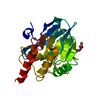
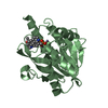
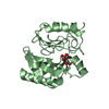
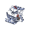
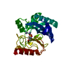
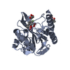
| Title | Crystal Structure of VIM-2 metallo-beta-lactamase in complex with ML302 and ML302F | ||||||
 Components Components | Beta-lactamase class B VIM-2 | ||||||
 Keywords Keywords | HYDROLASE/HYDROLASE INHIBITOR / alpha-beta/beta-alpha fold / beta-lactamase / HYDROLASE-HYDROLASE INHIBITOR complex | ||||||
| Function / homology |  Function and homology information Function and homology informationantibiotic catabolic process / beta-lactamase activity / beta-lactamase / periplasmic space / response to antibiotic / metal ion binding Similarity search - Function | ||||||
| Biological species |   Pseudomonas aeruginosa (bacteria) Pseudomonas aeruginosa (bacteria) | ||||||
| Method |  X-RAY DIFFRACTION / SYNCHROTRON / MOLECULAR REPLACEMENT / molecular replacement / Resolution: 1.48 Å X-RAY DIFFRACTION / SYNCHROTRON / MOLECULAR REPLACEMENT / molecular replacement / Resolution: 1.48 Å | ||||||
 Authors Authors | Aik, W.S. / Brem, J. / McDonough, M.A. / Schofield, C.J. | ||||||
 Citation Citation | Journal: Nat.Chem. / Year: 2014 Title: Rhodanine hydrolysis leads to potent thioenolate mediated metallo-beta-lactamase inhibition. Authors: Brem, J. / van Berkel, S.S. / Aik, W. / Rydzik, A.M. / Avison, M.B. / Pettinati, I. / Umland, K.D. / Kawamura, A. / Spencer, J. / Claridge, T.D. / McDonough, M.A. / Schofield, C.J. | ||||||
| History |
|
- Structure visualization
| Structure viewer | Molecule: MolmilJmol/JSmol |
|---|
- Downloads & links
-Download
| PDBx/mmCIF format | 4pvo.cif.gz | 201.4 KB | Display | PDBx/mmCIF format |
|---|---|---|---|---|
| PDB format | pdb4pvo.ent.gz | 158.6 KB | Display | PDB format |
| PDBx/mmJSON format | 4pvo.json.gz | Tree view | PDBx/mmJSON format | |
| Others |  Other downloads Other downloads |
-Validation report
| Arichive directory | https://data.pdbj.org/pub/pdb/validation_reports/pv/4pvoftp://data.pdbj.org/pub/pdb/validation_reports/pv/4pvo | HTTPS FTP |
|---|
-Related structure data
| Related structure data | 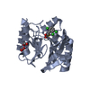 4pvtC 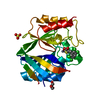 4tytC  1ko3S S: Starting model for refinement C: citing same article ( |
|---|---|
| Similar structure data |


-Links
 PDBj
PDBj

- Assembly
| Deposited unit | 
| ||||||||
|---|---|---|---|---|---|---|---|---|---|
| 1 | 
| ||||||||
| 2 | 
| ||||||||
| Unit cell |
|
-Components
-Protein , 1 types, 2 molecules AB
| #1: Protein | Mass: 25693.488 Da / Num. of mol.: 2 / Fragment: UNP RESIDUES 27-266 Source method: isolated from a genetically manipulated source Source: (gene. exp.) Pseudomonas aeruginosa (bacteria) / Gene: bla-VIM-2, blasVIM-2, blaVIM-2, blaVIM2, VIM-2 / Plasmid: pOPIN-F VIM-2 / Production host: |
|---|
-Non-polymers , 6 types, 550 molecules 


| #2: Chemical | ChemComp-ZN /  Mass: 65.409 Da / Num. of mol.: 6 / Source method: obtained synthetically / Formula: Zn Mass: 65.409 Da / Num. of mol.: 6 / Source method: obtained synthetically / Formula: Zn#3: Chemical |  Mass: 283.559 Da / Num. of mol.: 2 / Source method: obtained synthetically / Formula: C9H5Cl3O2S Mass: 283.559 Da / Num. of mol.: 2 / Source method: obtained synthetically / Formula: C9H5Cl3O2S#4: Chemical | ChemComp-SVB / |  Mass: 479.831 Da / Num. of mol.: 1 / Source method: obtained synthetically / Formula: C17H17Cl3N4O2S2 Mass: 479.831 Da / Num. of mol.: 1 / Source method: obtained synthetically / Formula: C17H17Cl3N4O2S2#5: Chemical | ChemComp-FMT /  Mass: 46.025 Da / Num. of mol.: 4 / Source method: obtained synthetically / Formula: CH2O2 Mass: 46.025 Da / Num. of mol.: 4 / Source method: obtained synthetically / Formula: CH2O2#6: Chemical | ChemComp-DMS / |  Mass: 78.133 Da / Num. of mol.: 1 / Source method: obtained synthetically / Formula: C2H6OS / Comment: DMSO, precipitant*YM Mass: 78.133 Da / Num. of mol.: 1 / Source method: obtained synthetically / Formula: C2H6OS / Comment: DMSO, precipitant*YM#7: Water | ChemComp-HOH / | Mass: 18.015 Da / Num. of mol.: 536 / Source method: isolated from a natural source / Formula: H2O |
|---|
-Experimental details
-Experiment
| Experiment | Method: X-RAY DIFFRACTION / Number of used crystals: 1 |
|---|
- Sample preparation
| Crystal | Density Matthews: 2.05 Å3/Da / Density % sol: 39.99 % / Mosaicity: 0.2 ° / Mosaicity esd: 0.004 ° |
|---|---|
| Crystal grow | Temperature: 293 K / Method: vapor diffusion, sitting drop / pH: 7.5 Details: 1mM TCEP, 2.5mM ML302, 50mM HEPES pH 7.5, 100mM NaCl, 0.1mM ZnCl2, 0.1M magnesium formate, 25% PEG 3350, VAPOR DIFFUSION, SITTING DROP, temperature 293K |
-Data collection
| Diffraction | Mean temperature: 100 K | |||||||||||||||||||||||||||||||||||||||||||||||||||||||||||||||||||||||||||||
|---|---|---|---|---|---|---|---|---|---|---|---|---|---|---|---|---|---|---|---|---|---|---|---|---|---|---|---|---|---|---|---|---|---|---|---|---|---|---|---|---|---|---|---|---|---|---|---|---|---|---|---|---|---|---|---|---|---|---|---|---|---|---|---|---|---|---|---|---|---|---|---|---|---|---|---|---|---|---|
| Diffraction source | Source: SYNCHROTRON / Site: Diamond  / Beamline: I04 / Wavelength: 0.8344 Å / Beamline: I04 / Wavelength: 0.8344 Å | |||||||||||||||||||||||||||||||||||||||||||||||||||||||||||||||||||||||||||||
| Detector | Type: PSI PILATUS 6M / Detector: PIXEL / Date: Sep 25, 2013 / Details: mirrors | |||||||||||||||||||||||||||||||||||||||||||||||||||||||||||||||||||||||||||||
| Radiation | Monochromator: double crystal / Protocol: SINGLE WAVELENGTH / Monochromatic (M) / Laue (L): M / Scattering type: x-ray | |||||||||||||||||||||||||||||||||||||||||||||||||||||||||||||||||||||||||||||
| Radiation wavelength | Wavelength: 0.8344 Å / Relative weight: 1 | |||||||||||||||||||||||||||||||||||||||||||||||||||||||||||||||||||||||||||||
| Reflection | Resolution: 1.48→50 Å / Num. all: 68860 / Num. obs: 68140 / % possible obs: 98.954 % / Observed criterion σ(F): -3 / Redundancy: 5 % / Biso Wilson estimate: 10.65 Å2 / Rmerge(I) obs: 0.136 / Χ2: 1.202 / Net I/σ(I): 13 | |||||||||||||||||||||||||||||||||||||||||||||||||||||||||||||||||||||||||||||
| Reflection shell | Diffraction-ID: 1 / Rejects: _
|
-Phasing
| Phasing | Method: molecular replacement |
|---|
- Processing
| Software |
| ||||||||||||||||||||||||||||||||||||
|---|---|---|---|---|---|---|---|---|---|---|---|---|---|---|---|---|---|---|---|---|---|---|---|---|---|---|---|---|---|---|---|---|---|---|---|---|---|
| Refinement | Method to determine structure: MOLECULAR REPLACEMENT Starting model: PDB ID 1KO3 Resolution: 1.48→39.1 Å / Isotropic thermal model: isotropic
| ||||||||||||||||||||||||||||||||||||
| Displacement parameters | Biso max: 70.96 Å2 / Biso mean: 17.7609 Å2 / Biso min: 5.05 Å2 | ||||||||||||||||||||||||||||||||||||
| Refinement step | Cycle: LAST / Resolution: 1.48→39.1 Å
| ||||||||||||||||||||||||||||||||||||
| Refine LS restraints |
| ||||||||||||||||||||||||||||||||||||
| LS refinement shell |
|
