 Movie
Movie Controller


+ Open data

- Basic information
| Entry | Database: PDB / ID: 4prs | ||||||
|---|---|---|---|---|---|---|---|
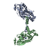
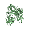
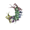
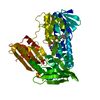
| Title | Structure of apo ArgBP from T. maritima | ||||||
 Components Components | ABC-type transporter, periplasmic subunit family 3 | ||||||
 Keywords Keywords | PROTEIN TRANSPORT / alpha/beta / Arginine binding | ||||||
| Function / homology |  Function and homology information Function and homology informationamino acid binding / ligand-gated monoatomic ion channel activity / outer membrane-bounded periplasmic space / membrane Similarity search - Function | ||||||
| Biological species |   Thermotoga maritima (bacteria) Thermotoga maritima (bacteria) | ||||||
| Method |  X-RAY DIFFRACTION / SYNCHROTRON / MAD / Resolution: 1.47 Å X-RAY DIFFRACTION / SYNCHROTRON / MAD / Resolution: 1.47 Å | ||||||
 Authors Authors | Ruggiero, A. / Dattelbaum, J.D. / Staiano, M. / Berisio, R. / D'Auria, S. / Vitagliano, L. | ||||||
 Citation Citation | Journal: Plos One / Year: 2014 Title: A loose domain swapping organization confers a remarkable stability to the dimeric structure of the arginine binding protein from Thermotoga maritima Authors: Ruggiero, A. / Dattelbaum, J.D. / Staiano, M. / Berisio, R. / D'Auria, S. / Vitagliano, L. | ||||||
| History |
|
- Structure visualization
| Structure viewer | Molecule: MolmilJmol/JSmol |
|---|
- Downloads & links
-Download
| PDBx/mmCIF format | 4prs.cif.gz | 218.3 KB | Display | PDBx/mmCIF format |
|---|---|---|---|---|
| PDB format | pdb4prs.ent.gz | 177.9 KB | Display | PDB format |
| PDBx/mmJSON format | 4prs.json.gz | Tree view | PDBx/mmJSON format | |
| Others |  Other downloads Other downloads |
-Validation report
| Arichive directory | https://data.pdbj.org/pub/pdb/validation_reports/pr/4prsftp://data.pdbj.org/pub/pdb/validation_reports/pr/4prs | HTTPS FTP |
|---|
-Related structure data


-Links
 PDBj
PDBj
- Assembly
| Deposited unit | 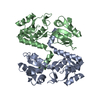
| |||||||||
|---|---|---|---|---|---|---|---|---|---|---|
| 1 |
| |||||||||
| Unit cell |
| |||||||||
| Components on special symmetry positions |
|
-Components
| #1: Protein | Mass: 25297.967 Da / Num. of mol.: 2 Source method: isolated from a genetically manipulated source Source: (gene. exp.) Thermotoga maritima (bacteria) / Strain: MSB8 / Gene: TM_0593 / Production host: #2: Water | ChemComp-HOH / |  Mass: 18.015 Da / Num. of mol.: 807 / Source method: isolated from a natural source / Formula: H2O Mass: 18.015 Da / Num. of mol.: 807 / Source method: isolated from a natural source / Formula: H2O |
|---|
-Experimental details
-Experiment
| Experiment | Method: X-RAY DIFFRACTION / Number of used crystals: 1 |
|---|
- Sample preparation
| Crystal | Density Matthews: 2.5 Å3/Da / Density % sol: 50.71 % |
|---|---|
| Crystal grow | Temperature: 293 K / Method: vapor diffusion, hanging drop / pH: 4.6 Details: 25%(w/v) PEG 3350, 0.1M Sodium Acetate trihydrate, pH 4.6, VAPOR DIFFUSION, HANGING DROP, temperature 293K |
-Data collection
| Diffraction | Mean temperature: 100 K | ||||||||||||
|---|---|---|---|---|---|---|---|---|---|---|---|---|---|
| Diffraction source | Source: SYNCHROTRON / Site: EMBL/DESY, HAMBURG  / Beamline: X12 / Wavelength: 0.9796, 0.9894, 0.9537 / Beamline: X12 / Wavelength: 0.9796, 0.9894, 0.9537 | ||||||||||||
| Detector | Type: MARMOSAIC 225 mm CCD / Detector: CCD | ||||||||||||
| Radiation | Protocol: MAD / Monochromatic (M) / Laue (L): M / Scattering type: x-ray | ||||||||||||
| Radiation wavelength |
| ||||||||||||
| Reflection | Resolution: 1.47→50 Å / Num. all: 86978 / Num. obs: 85761 / % possible obs: 98.7 % / Observed criterion σ(F): 0 / Observed criterion σ(I): 0 | ||||||||||||
| Reflection shell | Resolution: 1.47→1.52 Å / % possible all: 87.3 |
- Processing
| Software |
| ||||||||||||||||||||||||||||||||||||||||||||||||||||||||||||||||||||||||||||||||
|---|---|---|---|---|---|---|---|---|---|---|---|---|---|---|---|---|---|---|---|---|---|---|---|---|---|---|---|---|---|---|---|---|---|---|---|---|---|---|---|---|---|---|---|---|---|---|---|---|---|---|---|---|---|---|---|---|---|---|---|---|---|---|---|---|---|---|---|---|---|---|---|---|---|---|---|---|---|---|---|---|---|
| Refinement | Method to determine structure: MAD / Resolution: 1.47→14.82 Å / Cor.coef. Fo:Fc: 0.972 / Cor.coef. Fo:Fc free: 0.953 / SU B: 2.694 / SU ML: 0.045 / Cross valid method: THROUGHOUT / σ(F): 0 / ESU R: 0.08 / ESU R Free: 0.075 / Stereochemistry target values: MAXIMUM LIKELIHOOD / Details: HYDROGENS HAVE BEEN ADDED IN THE RIDING POSITIONS
| ||||||||||||||||||||||||||||||||||||||||||||||||||||||||||||||||||||||||||||||||
| Solvent computation | Ion probe radii: 0.8 Å / Shrinkage radii: 0.8 Å / VDW probe radii: 1.4 Å / Solvent model: BABINET MODEL WITH MASK | ||||||||||||||||||||||||||||||||||||||||||||||||||||||||||||||||||||||||||||||||
| Displacement parameters | Biso mean: 21.122 Å2
| ||||||||||||||||||||||||||||||||||||||||||||||||||||||||||||||||||||||||||||||||
| Refinement step | Cycle: LAST / Resolution: 1.47→14.82 Å
| ||||||||||||||||||||||||||||||||||||||||||||||||||||||||||||||||||||||||||||||||
| Refine LS restraints |
| ||||||||||||||||||||||||||||||||||||||||||||||||||||||||||||||||||||||||||||||||
| LS refinement shell | Resolution: 1.469→1.507 Å / Total num. of bins used: 20 /
|
