 Movie
Movie Controller

[English] 日本語
 Yorodumi
- PDB-4pm6: Crystal structure of CTX-M-14 S70G beta-lactamase at 1.56 Angstro... -
+ Open data

- Basic information
| Entry | Database: PDB / ID: 4pm6 | |||||||||
|---|---|---|---|---|---|---|---|---|---|---|
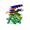
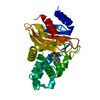
| Title | Crystal structure of CTX-M-14 S70G beta-lactamase at 1.56 Angstroms resolution | |||||||||
 Components Components | Beta-lactamase CTX-M-14 | |||||||||
 Keywords Keywords | HYDROLASE / class A beta-lactamase | |||||||||
| Function / homology |  Function and homology information Function and homology informationbeta-lactam antibiotic catabolic process / beta-lactamase activity / beta-lactamase / response to antibiotic Similarity search - Function | |||||||||
| Biological species |  Klebsiella pneumoniae subsp. pneumoniae (bacteria) Klebsiella pneumoniae subsp. pneumoniae (bacteria) | |||||||||
| Method |  X-RAY DIFFRACTION / MOLECULAR REPLACEMENT / Resolution: 1.56 Å X-RAY DIFFRACTION / MOLECULAR REPLACEMENT / Resolution: 1.56 Å | |||||||||
 Authors Authors | Adamski, C.J. / Cardenas, A.M. / Sankaran, B. / Palzkill, T. | |||||||||
| Funding support |  United States, 2items United States, 2items
| |||||||||
 Citation Citation | Journal: Biochemistry / Year: 2015 Title: Molecular Basis for the Catalytic Specificity of the CTX-M Extended-Spectrum beta-Lactamases. Authors: Adamski, C.J. / Cardenas, A.M. / Brown, N.G. / Horton, L.B. / Sankaran, B. / Prasad, B.V. / Gilbert, H.F. / Palzkill, T. | |||||||||
| History |
|
- Structure visualization
| Structure viewer | Molecule: MolmilJmol/JSmol |
|---|
- Downloads & links
-Download
| PDBx/mmCIF format | 4pm6.cif.gz | 73.5 KB | Display | PDBx/mmCIF format |
|---|---|---|---|---|
| PDB format | pdb4pm6.ent.gz | 51.3 KB | Display | PDB format |
| PDBx/mmJSON format | 4pm6.json.gz | Tree view | PDBx/mmJSON format | |
| Others |  Other downloads Other downloads |
-Validation report
| Arichive directory | https://data.pdbj.org/pub/pdb/validation_reports/pm/4pm6ftp://data.pdbj.org/pub/pdb/validation_reports/pm/4pm6 | HTTPS FTP |
|---|
-Related structure data
| Related structure data | 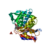 4pm5C 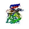 4pm7C 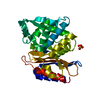 4pm8C 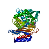 4pm9C 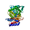 4pmaC  1yltS C: citing same article ( S: Starting model for refinement |
|---|---|
| Similar structure data |


-Links
 PDBj
PDBj


- Assembly
| Deposited unit | 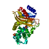
| ||||||||
|---|---|---|---|---|---|---|---|---|---|
| 1 |
| ||||||||
| Unit cell |
|
-Components
| #1: Protein | Mass: 27970.520 Da / Num. of mol.: 1 / Fragment: UNP residues 29-291 / Mutation: S70G Source method: isolated from a genetically manipulated source Source: (gene. exp.) Klebsiella pneumoniae subsp. pneumoniae (bacteria)Strain: HS11286 / Gene: KPHS_p301310 / Production host: | ||
|---|---|---|---|
| #2: Chemical |   Mass: 96.063 Da / Num. of mol.: 3 / Source method: obtained synthetically / Formula: SO4 Mass: 96.063 Da / Num. of mol.: 3 / Source method: obtained synthetically / Formula: SO4#3: Water | ChemComp-HOH / |  Mass: 18.015 Da / Num. of mol.: 395 / Source method: isolated from a natural source / Formula: H2O Mass: 18.015 Da / Num. of mol.: 395 / Source method: isolated from a natural source / Formula: H2O |
-Experimental details
-Experiment
| Experiment | Method: X-RAY DIFFRACTION / Number of used crystals: 1 |
|---|
- Sample preparation
| Crystal | Density Matthews: 1.99 Å3/Da / Density % sol: 38.31 % |
|---|---|
| Crystal grow | Temperature: 296 K / Method: vapor diffusion, hanging drop / Details: Ammonium Sulfate, PEG 4000 |
-Data collection
| Diffraction | Mean temperature: 80 K |
|---|---|
| Diffraction source | Source: ROTATING ANODE / Type: RIGAKU / Wavelength: 1.5 Å |
| Detector | Type: RIGAKU / Detector: IMAGE PLATE / Date: Apr 21, 2014 |
| Radiation | Protocol: SINGLE WAVELENGTH / Monochromatic (M) / Laue (L): M / Scattering type: x-ray |
| Radiation wavelength | Wavelength: 1.5 Å / Relative weight: 1 |
| Reflection | Resolution: 1.56→22.1 Å / Num. obs: 32576 / % possible obs: 100 % / Redundancy: 2 % / Net I/σ(I): 39.4 |
- Processing
| Software | Name: PHENIX / Version: (phenix.refine: 1.8.4_1496) / Classification: refinement | |||||||||||||||||||||||||||||||||||||||||||||||||||||||||||||||||||||||||||||||||||||||||||||||||||||||||||||||||||||||||||||||||||||||||||||||||||||||||||||||||
|---|---|---|---|---|---|---|---|---|---|---|---|---|---|---|---|---|---|---|---|---|---|---|---|---|---|---|---|---|---|---|---|---|---|---|---|---|---|---|---|---|---|---|---|---|---|---|---|---|---|---|---|---|---|---|---|---|---|---|---|---|---|---|---|---|---|---|---|---|---|---|---|---|---|---|---|---|---|---|---|---|---|---|---|---|---|---|---|---|---|---|---|---|---|---|---|---|---|---|---|---|---|---|---|---|---|---|---|---|---|---|---|---|---|---|---|---|---|---|---|---|---|---|---|---|---|---|---|---|---|---|---|---|---|---|---|---|---|---|---|---|---|---|---|---|---|---|---|---|---|---|---|---|---|---|---|---|---|---|---|---|---|---|
| Refinement | Method to determine structure: MOLECULAR REPLACEMENT Starting model: 1YLT Resolution: 1.56→22.096 Å / SU ML: 0.12 / Cross valid method: NONE / σ(F): 1.96 / Phase error: 14.75 / Stereochemistry target values: ML
| |||||||||||||||||||||||||||||||||||||||||||||||||||||||||||||||||||||||||||||||||||||||||||||||||||||||||||||||||||||||||||||||||||||||||||||||||||||||||||||||||
| Solvent computation | Shrinkage radii: 0.9 Å / VDW probe radii: 1.11 Å / Solvent model: FLAT BULK SOLVENT MODEL | |||||||||||||||||||||||||||||||||||||||||||||||||||||||||||||||||||||||||||||||||||||||||||||||||||||||||||||||||||||||||||||||||||||||||||||||||||||||||||||||||
| Refinement step | Cycle: LAST / Resolution: 1.56→22.096 Å
| |||||||||||||||||||||||||||||||||||||||||||||||||||||||||||||||||||||||||||||||||||||||||||||||||||||||||||||||||||||||||||||||||||||||||||||||||||||||||||||||||
| Refine LS restraints |
| |||||||||||||||||||||||||||||||||||||||||||||||||||||||||||||||||||||||||||||||||||||||||||||||||||||||||||||||||||||||||||||||||||||||||||||||||||||||||||||||||
| LS refinement shell |
|
