 Movie
Movie Controller


+ Open data

- Basic information
| Entry | Database: PDB / ID: 1hzo | ||||||
|---|---|---|---|---|---|---|---|
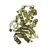
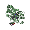
| Title | STRUCTURE OF CLASS A CEPHALOSPORINASE FROM PROTEUS VULGARIS K1 | ||||||
 Components Components | BETA-LACTAMASE | ||||||
 Keywords Keywords | HYDROLASE / MIXED ALPHA/BETA / CEPHALOSPORINASE / CLASS A BETA-LACTAMASE | ||||||
| Function / homology |  Function and homology information Function and homology informationbeta-lactam antibiotic catabolic process / beta-lactamase activity / beta-lactamase / response to antibiotic Similarity search - Function | ||||||
| Biological species |  Proteus vulgaris (bacteria) Proteus vulgaris (bacteria) | ||||||
| Method |  X-RAY DIFFRACTION / MOLECULAR REPLACEMENT / Resolution: 1.75 Å X-RAY DIFFRACTION / MOLECULAR REPLACEMENT / Resolution: 1.75 Å | ||||||
 Authors Authors | Nukaga, M. / Crichlow, G.V. / Kuzin, A.P. / Mayama, K. / Knox, J.R. | ||||||
 Citation Citation | Journal: J.Mol.Biol. / Year: 2002 Title: Structure of an extended-spectrum class A beta-lactamase from Proteus vulgaris K1. Authors: Nukaga, M. / Mayama, K. / Crichlow, G.V. / Knox, J.R. | ||||||
| History |
| ||||||
| Remark 999 | SEQUENCE THE RESIDUE NUMBERING IS NOT SEQUENTIAL. Residues 57 and 59, 238 and 240, 252 and 254 are ...SEQUENCE THE RESIDUE NUMBERING IS NOT SEQUENTIAL. Residues 57 and 59, 238 and 240, 252 and 254 are covalently bound. The numbering is the accepted consensus numbering scheme for beta-lactamases. Asp24-asn25-asn26 are not seen in the density at the N-terminus, and the last four residues are not seen at the C-term in both molecules. |
- Structure visualization
| Structure viewer | Molecule: MolmilJmol/JSmol |
|---|
- Downloads & links
-Download
| PDBx/mmCIF format | 1hzo.cif.gz | 128.6 KB | Display | PDBx/mmCIF format |
|---|---|---|---|---|
| PDB format | pdb1hzo.ent.gz | 98.8 KB | Display | PDB format |
| PDBx/mmJSON format | 1hzo.json.gz | Tree view | PDBx/mmJSON format | |
| Others |  Other downloads Other downloads |
-Validation report
| Arichive directory | https://data.pdbj.org/pub/pdb/validation_reports/hz/1hzoftp://data.pdbj.org/pub/pdb/validation_reports/hz/1hzo | HTTPS FTP |
|---|
-Related structure data
| Related structure data |  1bzaS S: Starting model for refinement |
|---|---|
| Similar structure data |


-Links
 PDBj
PDBj


- Assembly
| Deposited unit | 
| ||||||||
|---|---|---|---|---|---|---|---|---|---|
| 1 | 
| ||||||||
| 2 | 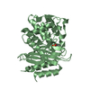
| ||||||||
| Unit cell |
|
-Components
| #1: Protein | Mass: 29689.396 Da / Num. of mol.: 2 Source method: isolated from a genetically manipulated source Source: (gene. exp.) Proteus vulgaris (bacteria) / Strain: K1 / Gene: BLAC / Plasmid: PPVCF1 / Production host: #2: Chemical |   Mass: 195.237 Da / Num. of mol.: 2 / Source method: obtained synthetically / Formula: C6H13NO4S / Comment: pH buffer*YM Mass: 195.237 Da / Num. of mol.: 2 / Source method: obtained synthetically / Formula: C6H13NO4S / Comment: pH buffer*YM#3: Water | ChemComp-HOH / |  Mass: 18.015 Da / Num. of mol.: 663 / Source method: isolated from a natural source / Formula: H2O Mass: 18.015 Da / Num. of mol.: 663 / Source method: isolated from a natural source / Formula: H2O |
|---|
-Experimental details
-Experiment
| Experiment | Method: X-RAY DIFFRACTION / Number of used crystals: 1 |
|---|
- Sample preparation
| Crystal | Density Matthews: 2.2 Å3/Da / Density % sol: 44 % | ||||||||||||||||||||||||||||||||||||
|---|---|---|---|---|---|---|---|---|---|---|---|---|---|---|---|---|---|---|---|---|---|---|---|---|---|---|---|---|---|---|---|---|---|---|---|---|---|
| Crystal grow | Temperature: 293 K / Method: vapor diffusion, sitting drop / pH: 6.25 Details: PEG 6000, MES, pH 6.25, VAPOR DIFFUSION, SITTING DROP, temperature 293K | ||||||||||||||||||||||||||||||||||||
| Crystal | *PLUS Density % sol: 44.1 % | ||||||||||||||||||||||||||||||||||||
| Crystal grow | *PLUS | ||||||||||||||||||||||||||||||||||||
| Components of the solutions | *PLUS
|
-Data collection
| Diffraction | Mean temperature: 100 K |
|---|---|
| Diffraction source | Source: ROTATING ANODE / Type: RIGAKU RU200 / Wavelength: 1.542 Å |
| Detector | Type: SIEMENS HI-STAR / Detector: AREA DETECTOR / Date: May 3, 1999 / Details: Franks mirrors |
| Radiation | Monochromator: Ni FILTER / Protocol: SINGLE WAVELENGTH / Monochromatic (M) / Laue (L): M / Scattering type: x-ray |
| Radiation wavelength | Wavelength: 1.542 Å / Relative weight: 1 |
| Reflection | Resolution: 1.75→44 Å / Num. all: 53765 / Num. obs: 49364 / % possible obs: 91.8 % / Observed criterion σ(F): 0 / Observed criterion σ(I): 0 / Redundancy: 5.76 % / Biso Wilson estimate: 24 Å2 / Rsym value: 5.2 / Net I/σ(I): 18.5 |
| Reflection shell | Resolution: 1.75→1.86 Å / Redundancy: 2.7 % / Mean I/σ(I) obs: 3.2 / Num. unique all: 5658 / Rsym value: 20.3 / % possible all: 64.6 |
| Reflection | *PLUS % possible obs: 92 % / Num. measured all: 284238 / Rmerge(I) obs: 0.052 |
| Reflection shell | *PLUS % possible obs: 65 % / Num. unique obs: 5658 / Num. measured obs: 15124 / Rmerge(I) obs: 0.2 |
- Processing
| Software |
| |||||||||||||||||||||||||
|---|---|---|---|---|---|---|---|---|---|---|---|---|---|---|---|---|---|---|---|---|---|---|---|---|---|---|
| Refinement | Method to determine structure: MOLECULAR REPLACEMENT Starting model: PDB ENTRY 1BZA Resolution: 1.75→44 Å / Cross valid method: THROUGHOUT / σ(F): 0 / Stereochemistry target values: Engh & Huber
| |||||||||||||||||||||||||
| Solvent computation | Solvent model: flat model / Bsol: 42.2 Å2 / ksol: 0.32 e/Å3 | |||||||||||||||||||||||||
| Displacement parameters | Biso mean: 13.1 Å2 | |||||||||||||||||||||||||
| Refine analyze | Luzzati coordinate error obs: 0.17 Å / Luzzati d res low obs: 5 Å | |||||||||||||||||||||||||
| Refinement step | Cycle: LAST / Resolution: 1.75→44 Å
| |||||||||||||||||||||||||
| Refine LS restraints |
| |||||||||||||||||||||||||
| LS refinement shell | Resolution: 1.75→1.81 Å
| |||||||||||||||||||||||||
| Refinement | *PLUS Rfactor obs: 0.169 / Rfactor Rfree: 0.193 / Rfactor Rwork: 0.169 | |||||||||||||||||||||||||
| Solvent computation | *PLUS | |||||||||||||||||||||||||
| Displacement parameters | *PLUS | |||||||||||||||||||||||||
| Refine LS restraints | *PLUS
| |||||||||||||||||||||||||
| LS refinement shell | *PLUS Rfactor Rfree: 0.267 / Rfactor Rwork: 0.239 / Num. reflection obs: 2580 / Rfactor obs: 0.239 |
