 Movie
Movie Controller

[English] 日本語
 Yorodumi
- PDB-4pbv: Crystal structure of chicken receptor protein tyrosine phosphatas... -
+ Open data

- Basic information
| Entry | Database: PDB / ID: 4pbv | ||||||||||||||||||
|---|---|---|---|---|---|---|---|---|---|---|---|---|---|---|---|---|---|---|---|
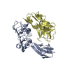
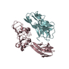
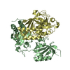
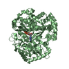
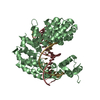
| Title | Crystal structure of chicken receptor protein tyrosine phosphatase sigma in complex with TrkC | ||||||||||||||||||
 Components Components |
| ||||||||||||||||||
 Keywords Keywords | SIGNALING PROTEIN / Synapse Cell signalling Cell surface receptor | ||||||||||||||||||
| Function / homology |  Function and homology information Function and homology informationReceptor-type tyrosine-protein phosphatases / NTF3 activates NTRK3 signaling / Activated NTRK3 signals through PLCG1 / negative regulation of collateral sprouting / negative regulation of axon regeneration / neurotrophin receptor activity / neurotrophin binding / negative regulation of dendritic spine development / synaptic membrane adhesion / negative regulation of axon extension ...Receptor-type tyrosine-protein phosphatases / NTF3 activates NTRK3 signaling / Activated NTRK3 signals through PLCG1 / negative regulation of collateral sprouting / negative regulation of axon regeneration / neurotrophin receptor activity / neurotrophin binding / negative regulation of dendritic spine development / synaptic membrane adhesion / negative regulation of axon extension / PIP3 activates AKT signaling / PI5P, PP2A and IER3 Regulate PI3K/AKT Signaling / Activated NTRK3 signals through PI3K / heparan sulfate proteoglycan binding / peptidyl-tyrosine dephosphorylation / protein-tyrosine-phosphatase / transmembrane receptor protein tyrosine kinase activity / protein tyrosine phosphatase activity / cell surface receptor protein tyrosine kinase signaling pathway / positive regulation of neuron projection development / receptor protein-tyrosine kinase / cellular response to nerve growth factor stimulus / postsynaptic density membrane / synaptic vesicle membrane / nervous system development / heparin binding / heart development / growth cone / perikaryon / cell differentiation / positive regulation of phosphatidylinositol 3-kinase/protein kinase B signal transduction / receptor complex / axon / signal transduction / protein homodimerization activity / ATP binding / plasma membrane Similarity search - Function | ||||||||||||||||||
| Biological species |  | ||||||||||||||||||
| Method |  X-RAY DIFFRACTION / SYNCHROTRON / MOLECULAR REPLACEMENT / Resolution: 2.5 Å X-RAY DIFFRACTION / SYNCHROTRON / MOLECULAR REPLACEMENT / Resolution: 2.5 Å | ||||||||||||||||||
 Authors Authors | Coles, C.H. / Mitakidis, N. / Zhang, P. / Elegheert, J. / Lu, W. / Stoker, A.W. / Nakagawa, T. / Craig, A.M. / Jones, E.Y. / Aricescu, A.R. | ||||||||||||||||||
| Funding support |  United Kingdom, 5items United Kingdom, 5items
| ||||||||||||||||||
 Citation Citation | Journal: Nat Commun / Year: 2014 Title: Structural basis for extracellular cis and trans RPTP sigma signal competition in synaptogenesis. Authors: Coles, C.H. / Mitakidis, N. / Zhang, P. / Elegheert, J. / Lu, W. / Stoker, A.W. / Nakagawa, T. / Craig, A.M. / Jones, E.Y. / Aricescu, A.R. | ||||||||||||||||||
| History |
|
- Structure visualization
| Structure viewer | Molecule: MolmilJmol/JSmol |
|---|
- Downloads & links
-Download
| PDBx/mmCIF format | 4pbv.cif.gz | 445.8 KB | Display | PDBx/mmCIF format |
|---|---|---|---|---|
| PDB format | pdb4pbv.ent.gz | 364.6 KB | Display | PDB format |
| PDBx/mmJSON format | 4pbv.json.gz | Tree view | PDBx/mmJSON format | |
| Others |  Other downloads Other downloads |
-Validation report
| Summary document | 4pbv_validation.pdf.gz | 506.5 KB | Display | wwPDB validaton report |
|---|---|---|---|---|
| Full document | 4pbv_full_validation.pdf.gz | 510.9 KB | Display | |
| Data in XML | 4pbv_validation.xml.gz | 38.3 KB | Display | |
| Data in CIF | 4pbv_validation.cif.gz | 53.5 KB | Display | |
| Arichive directory | https://data.pdbj.org/pub/pdb/validation_reports/pb/4pbvftp://data.pdbj.org/pub/pdb/validation_reports/pb/4pbv | HTTPS FTP |
-Related structure data
| Related structure data | 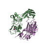 4pbwC  4pbxC  2yd4S S: Starting model for refinement C: citing same article ( |
|---|---|
| Similar structure data |


-Links
 PDBj
PDBj


- Assembly
| Deposited unit | 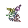
| ||||||||||||||||||||||||||||||||||||||||||||||||||||||||||||||||||||||||||||||||||||||||||||||||||||||||||||||||||||||||||
|---|---|---|---|---|---|---|---|---|---|---|---|---|---|---|---|---|---|---|---|---|---|---|---|---|---|---|---|---|---|---|---|---|---|---|---|---|---|---|---|---|---|---|---|---|---|---|---|---|---|---|---|---|---|---|---|---|---|---|---|---|---|---|---|---|---|---|---|---|---|---|---|---|---|---|---|---|---|---|---|---|---|---|---|---|---|---|---|---|---|---|---|---|---|---|---|---|---|---|---|---|---|---|---|---|---|---|---|---|---|---|---|---|---|---|---|---|---|---|---|---|---|---|---|
| 1 | 
| ||||||||||||||||||||||||||||||||||||||||||||||||||||||||||||||||||||||||||||||||||||||||||||||||||||||||||||||||||||||||||
| 2 | 
| ||||||||||||||||||||||||||||||||||||||||||||||||||||||||||||||||||||||||||||||||||||||||||||||||||||||||||||||||||||||||||
| 3 | 
| ||||||||||||||||||||||||||||||||||||||||||||||||||||||||||||||||||||||||||||||||||||||||||||||||||||||||||||||||||||||||||
| Unit cell |
| ||||||||||||||||||||||||||||||||||||||||||||||||||||||||||||||||||||||||||||||||||||||||||||||||||||||||||||||||||||||||||
| Noncrystallographic symmetry (NCS) | NCS domain:
NCS domain segments: Component-ID: _ / Refine code: _
NCS ensembles :
|
-Components
| #1: Protein | Mass: 30399.387 Da / Num. of mol.: 2 / Fragment: Residues 31-302 / Mutation: N163Q, N232Q, N259Q, N267Q and N294Q Source method: isolated from a genetically manipulated source Source: (gene. exp.)  Homo sapiens (human) Homo sapiens (human)References: UniProt: Q91044, receptor protein-tyrosine kinase #2: Protein | Mass: 32721.014 Da / Num. of mol.: 3 / Fragment: Residues 29-316 Source method: isolated from a genetically manipulated source Details: Ig3 domain is not visible in electron density, suggesting that this domain had either been proteolytically cleaved during crystallisation or is disordered. Source: (gene. exp.) Homo sapiens (human) / References: UniProt: Q90815, UniProt: F1NWE3*PLUS#3: Sugar | ChemComp-NAG /   Type: D-saccharide, beta linking / Mass: 221.208 Da / Num. of mol.: 5 / Source method: obtained synthetically / Formula: C8H15NO6 Type: D-saccharide, beta linking / Mass: 221.208 Da / Num. of mol.: 5 / Source method: obtained synthetically / Formula: C8H15NO6#4: Chemical |   Mass: 96.063 Da / Num. of mol.: 2 / Source method: obtained synthetically / Formula: SO4 Mass: 96.063 Da / Num. of mol.: 2 / Source method: obtained synthetically / Formula: SO4#5: Water | ChemComp-HOH / |  Mass: 18.015 Da / Num. of mol.: 118 / Source method: isolated from a natural source / Formula: H2O Mass: 18.015 Da / Num. of mol.: 118 / Source method: isolated from a natural source / Formula: H2OHas protein modification | Y | |
|---|
-Experimental details
-Experiment
| Experiment | Method: X-RAY DIFFRACTION |
|---|
- Sample preparation
| Crystal | Density Matthews: 2.44 Å3/Da / Density % sol: 49.49 % |
|---|---|
| Crystal grow | Temperature: 293.5 K / Method: vapor diffusion, sitting drop / pH: 8.5 Details: 10% PEG 20k, 20% PEG MME 550, 0.1M bicine/Tris pH8.5, 0.03M sodium nitrate, 0.03M disodium hydrogen phosphate, 0.03M ammonium sulphate |
-Data collection
| Diffraction | Mean temperature: 100 K |
|---|---|
| Diffraction source | Source: SYNCHROTRON / Site: Diamond / Beamline: I04 / Wavelength: 0.92 Å |
| Detector | Type: DECTRIS PILATUS 2M / Detector: PIXEL / Date: Aug 3, 2012 |
| Radiation | Protocol: SINGLE WAVELENGTH / Monochromatic (M) / Laue (L): M / Scattering type: x-ray |
| Radiation wavelength | Wavelength: 0.92 Å / Relative weight: 1 |
| Reflection | Resolution: 2.5→92.79 Å / Num. obs: 52662 / % possible obs: 99.8 % / Redundancy: 11.6 % / Net I/σ(I): 10.7 |
| Reflection shell | Resolution: 2.5→2.56 Å / Redundancy: 7.1 % / Mean I/σ(I) obs: 1.6 / % possible all: 99.3 |
- Processing
| Software |
| ||||||||||||||||||||||||||||||||||||||||||||||||||||||||||||||||||||||||||||||||||||||||||||||||||||||||||||||||||||||||||||||||||||||||||||||||||||||||||||||||||||||||||||||||||||||
|---|---|---|---|---|---|---|---|---|---|---|---|---|---|---|---|---|---|---|---|---|---|---|---|---|---|---|---|---|---|---|---|---|---|---|---|---|---|---|---|---|---|---|---|---|---|---|---|---|---|---|---|---|---|---|---|---|---|---|---|---|---|---|---|---|---|---|---|---|---|---|---|---|---|---|---|---|---|---|---|---|---|---|---|---|---|---|---|---|---|---|---|---|---|---|---|---|---|---|---|---|---|---|---|---|---|---|---|---|---|---|---|---|---|---|---|---|---|---|---|---|---|---|---|---|---|---|---|---|---|---|---|---|---|---|---|---|---|---|---|---|---|---|---|---|---|---|---|---|---|---|---|---|---|---|---|---|---|---|---|---|---|---|---|---|---|---|---|---|---|---|---|---|---|---|---|---|---|---|---|---|---|---|---|
| Refinement | Method to determine structure: MOLECULAR REPLACEMENT Starting model: 2YD4 Resolution: 2.5→92.79 Å / Cor.coef. Fo:Fc: 0.943 / Cor.coef. Fo:Fc free: 0.918 / SU B: 20.34 / SU ML: 0.213 / Cross valid method: FREE R-VALUE / ESU R: 0.405 / ESU R Free: 0.263 / Stereochemistry target values: MAXIMUM LIKELIHOOD / Details: HYDROGENS HAVE BEEN ADDED IN THE RIDING POSITIONS
| ||||||||||||||||||||||||||||||||||||||||||||||||||||||||||||||||||||||||||||||||||||||||||||||||||||||||||||||||||||||||||||||||||||||||||||||||||||||||||||||||||||||||||||||||||||||
| Solvent computation | Ion probe radii: 0.8 Å / Shrinkage radii: 0.8 Å / VDW probe radii: 1.2 Å / Solvent model: MASK | ||||||||||||||||||||||||||||||||||||||||||||||||||||||||||||||||||||||||||||||||||||||||||||||||||||||||||||||||||||||||||||||||||||||||||||||||||||||||||||||||||||||||||||||||||||||
| Displacement parameters | Biso mean: 56.72 Å2
| ||||||||||||||||||||||||||||||||||||||||||||||||||||||||||||||||||||||||||||||||||||||||||||||||||||||||||||||||||||||||||||||||||||||||||||||||||||||||||||||||||||||||||||||||||||||
| Refinement step | Cycle: 1 / Resolution: 2.5→92.79 Å
| ||||||||||||||||||||||||||||||||||||||||||||||||||||||||||||||||||||||||||||||||||||||||||||||||||||||||||||||||||||||||||||||||||||||||||||||||||||||||||||||||||||||||||||||||||||||
| Refine LS restraints |
|