 Movie
Movie Controller


+ Open data

- Basic information
| Entry | Database: PDB / ID: 4p7d | ||||||
|---|---|---|---|---|---|---|---|
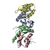
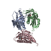
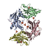
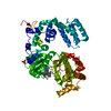
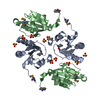
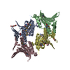
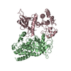
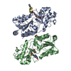
| Title | Antitoxin HicB3 crystal structure | ||||||
 Components Components | Antitoxin HicB3 | ||||||
 Keywords Keywords | TOXIN / toxin-antitoxin complex / homotetramer | ||||||
| Function / homology |  Function and homology information Function and homology informationsequence-specific DNA binding / regulation of DNA-templated transcription Similarity search - Function | ||||||
| Biological species |   Yersinia pestis (bacteria) Yersinia pestis (bacteria) | ||||||
| Method |  X-RAY DIFFRACTION / SYNCHROTRON / SAD / Resolution: 2.781 Å X-RAY DIFFRACTION / SYNCHROTRON / SAD / Resolution: 2.781 Å | ||||||
 Authors Authors | Li de la Sierra-Gallay, I. / Bibi-Triki, S. / van Tilbeurgh, H. / Lazar, N. / Pradel, E. | ||||||
 Citation Citation | Journal: J.Bacteriol. / Year: 2014 Title: Functional and Structural Analysis of HicA3-HicB3, a Novel Toxin-Antitoxin System of Yersinia pestis. Authors: Bibi-Triki, S. / Li de la Sierra-Gallay, I. / Lazar, N. / Leroy, A. / Van Tilbeurgh, H. / Sebbane, F. / Pradel, E. | ||||||
| History |
|
- Structure visualization
| Structure viewer | Molecule: MolmilJmol/JSmol |
|---|
- Downloads & links
-Download
| PDBx/mmCIF format | 4p7d.cif.gz | 117.8 KB | Display | PDBx/mmCIF format |
|---|---|---|---|---|
| PDB format | pdb4p7d.ent.gz | 93.4 KB | Display | PDB format |
| PDBx/mmJSON format | 4p7d.json.gz | Tree view | PDBx/mmJSON format | |
| Others |  Other downloads Other downloads |
-Validation report
| Arichive directory | https://data.pdbj.org/pub/pdb/validation_reports/p7/4p7dftp://data.pdbj.org/pub/pdb/validation_reports/p7/4p7d | HTTPS FTP |
|---|
-Related structure data


-Links
 PDBj
PDBj- Assembly
| Deposited unit | 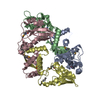
| ||||||||
|---|---|---|---|---|---|---|---|---|---|
| 1 |
| ||||||||
| Unit cell |
|
-Components
| #1: Protein | Mass: 16475.211 Da / Num. of mol.: 4 Source method: isolated from a genetically manipulated source Source: (gene. exp.) Yersinia pestis (bacteria) / Gene: YPO3369 / Production host: #2: Chemical |   Mass: 35.453 Da / Num. of mol.: 2 / Source method: obtained synthetically / Formula: Cl Mass: 35.453 Da / Num. of mol.: 2 / Source method: obtained synthetically / Formula: ClHas protein modification | Y | |
|---|
-Experimental details
-Experiment
| Experiment | Method: X-RAY DIFFRACTION |
|---|
- Sample preparation
| Crystal | Density Matthews: 2.41 Å3/Da / Density % sol: 48.89 % |
|---|---|
| Crystal grow | Temperature: 293 K / Method: vapor diffusion, hanging drop / pH: 6.5 / Details: polyethylene glycol 3000, MES pH 6.5 |
-Data collection
| Diffraction | Mean temperature: 100 K |
|---|---|
| Diffraction source | Source: SYNCHROTRON / Site: SOLEIL  / Beamline: PROXIMA 1 / Wavelength: 0.97911 Å / Beamline: PROXIMA 1 / Wavelength: 0.97911 Å |
| Detector | Type: PSI PILATUS 6M / Detector: PIXEL / Date: Dec 5, 2012 |
| Radiation | Protocol: SINGLE WAVELENGTH / Monochromatic (M) / Laue (L): M / Scattering type: x-ray |
| Radiation wavelength | Wavelength: 0.97911 Å / Relative weight: 1 |
| Reflection | Resolution: 2.78→46.85 Å / Num. obs: 16491 / % possible obs: 99.5 % / Redundancy: 7.1 % / Biso Wilson estimate: 69.76 Å2 / Rmerge(I) obs: 0.095 / Net I/σ(I): 14.45 |
| Reflection shell | Resolution: 2.78→2.95 Å / Redundancy: 7 % / Rmerge(I) obs: 0.683 / Mean I/σ(I) obs: 2.84 / % possible all: 97.2 |
- Processing
| Software |
| |||||||||||||||||||||||||||||||||||||||||||||||||
|---|---|---|---|---|---|---|---|---|---|---|---|---|---|---|---|---|---|---|---|---|---|---|---|---|---|---|---|---|---|---|---|---|---|---|---|---|---|---|---|---|---|---|---|---|---|---|---|---|---|---|
| Refinement | Method to determine structure: SAD / Resolution: 2.781→46.846 Å / SU ML: 0.42 / Cross valid method: FREE R-VALUE / σ(F): 1.36 / Phase error: 30.39 / Stereochemistry target values: ML
| |||||||||||||||||||||||||||||||||||||||||||||||||
| Solvent computation | Shrinkage radii: 0.9 Å / VDW probe radii: 1.11 Å / Solvent model: FLAT BULK SOLVENT MODEL | |||||||||||||||||||||||||||||||||||||||||||||||||
| Displacement parameters | Biso mean: 63.42 Å2 | |||||||||||||||||||||||||||||||||||||||||||||||||
| Refinement step | Cycle: LAST / Resolution: 2.781→46.846 Å
| |||||||||||||||||||||||||||||||||||||||||||||||||
| Refine LS restraints |
| |||||||||||||||||||||||||||||||||||||||||||||||||
| LS refinement shell |
|
